Amoeba cell under microscope
Home » Science Education » Amoeba cell under microscopeAmoeba cell under microscope
Amoeba Cell Under Microscope. Stentor eats arcella youtube. Onion cells and cheek cells my science blog. Amoeba under the microscope amoeba is a unicellular organism in the kingdom protozoa. Amoebas are usually considered among the lowest and most primitive forms of life.
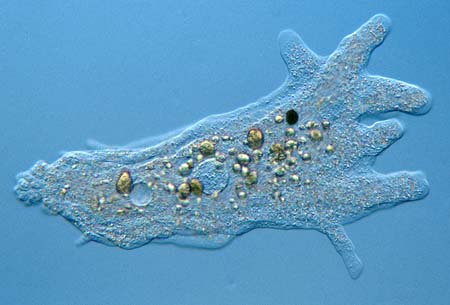 Mic Uk Amoebas Are More Than Just Blobs From microscopy-uk.org.uk
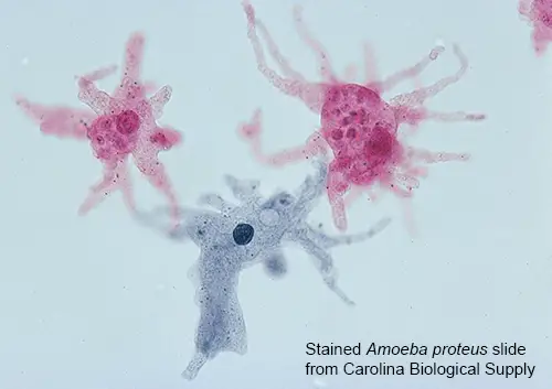
These microscopic organisms are often called shape shifters as they have the ability to constantly change their shape. The top image of amoeba proteus gives a good insight in the amoeba s anatomy. Amoebas have a single cell that appears to be not much more than cytoplasm held together by a flexible cell wall. Amoeba under the microscope fixing staining techniques and structure amoeba plural amoebas amoebae is a genus that belongs to kingdom protozoa. Generally the term is used to describe single celled organisms that move in a primitive crawling manner by using temporary false feet known as pseudopods. Amoeba under microscope amoeba are shapeless they look like a big blob unicellular organisms from the genus protozoa.
Amoeba under microscope amoeba are shapeless they look like a big blob unicellular organisms from the genus protozoa.
These microscopic organisms are often called shape shifters as they have the ability to constantly change their shape. Amoeba under microscope amoeba are shapeless they look like a big blob unicellular organisms from the genus protozoa. Balamuthia mandrillaris is the cause of often fatal granulomatous amoebic meningoencephalitis. A tiny blob of colorless jelly with a dark speck inside it this is what an amoeba looks like when seen through a microscope. Micro 2210 study guide 2013 14 vito instructor vito at. Amoeba under the microscope amoeba is a unicellular organism in the kingdom protozoa.
 Source: davidwangblog.wordpress.com
A tiny blob of colorless jelly with a dark speck inside it this is what an amoeba looks like when seen through a microscope. Micro 2210 study guide 2013 14 vito instructor vito at. Boundary driven oscillations rescue pdsa cells biorxiv. Amoebas have a single cell that appears to be not much more than cytoplasm held together by a flexible cell wall. Balamuthia mandrillaris is the cause of often fatal granulomatous amoebic meningoencephalitis.
 Source: blog.microscopeworld.com
Amoebae can likewise play host to microscopic organisms that are pathogenic to people and help in spreading such microbes. A tiny blob of colorless jelly with a dark speck inside it this is what an amoeba looks like when seen through a microscope. Floating in this cytoplasm all kinds of cell bodies can be found. Amoebas have a single cell that appears to be not much more than cytoplasm held together by a flexible cell wall. Onion cells and cheek cells my science blog.
Source: enchantedlearning.com
Amoeba is an aquatic single cell unicellular organism with membrane bound eukaryotic organelles that has no definite shape. Onion cells and cheek cells my science blog. Image courtesy pearson scott foresman. Amoeba is an aquatic single cell unicellular organism with membrane bound eukaryotic organelles that has no definite shape. When seen under a microscope the cell looks like a tiny blob of colorless jelly with a dark speck inside it.
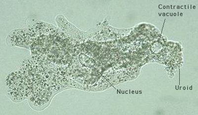 Source: daviddarling.info
Microscopy of nosema for beekeepers. Micro 2210 study guide 2013 14 vito instructor vito at. Amoeba under the microscope amoeba is a unicellular organism in the kingdom protozoa. These microscopic organisms are often called shape shifters as they have the ability to constantly change their shape. Amoeba under the microscope fixing staining techniques and structure amoeba plural amoebas amoebae is a genus that belongs to kingdom protozoa.
 Source: youtube.com
Balamuthia mandrillaris is the cause of often fatal granulomatous amoebic meningoencephalitis. The colorless jelly is cytoplasm and the dark speck is the nucleus. Amoeba under the microscope fixing staining techniques structure. It is capable of movement. Micro 2210 study guide 2013 14 vito instructor vito at.
 Source: m.youtube.com
Amoebas have a single cell that appears to be not much more than cytoplasm held together by a flexible cell wall. The most obvious is the nucleus. The colorless jelly is cytoplasm and the dark speck is the nucleus. Generally the term is used to describe single celled organisms that move in a primitive crawling manner by using temporary false feet known as pseudopods. Amoeba under microscope amoeba are shapeless they look like a big blob unicellular organisms from the genus protozoa.
 Source: britannica.com
Amoeba moves with their pseudopodia which are a specialized form of the plasma membrane that results in a crawling motion of the organism. The most obvious is the nucleus. Amoeba under the microscope fixing staining techniques and structure amoeba plural amoebas amoebae is a genus that belongs to kingdom protozoa. Amoeba under microscope amoeba are shapeless they look like a big blob unicellular organisms from the genus protozoa. Amoeba is an aquatic single cell unicellular organism with membrane bound eukaryotic organelles that has no definite shape.
 Source: amazon.com
The term amoeba is from the greek word amoibe which means to change. Balamuthia mandrillaris is the cause of often fatal granulomatous amoebic meningoencephalitis. Image courtesy pearson scott foresman. Amoebae can likewise play host to microscopic organisms that are pathogenic to people and help in spreading such microbes. Amoebas have a single cell that appears to be not much more than cytoplasm held together by a flexible cell wall.
 Source: microscopemaster.com
Amoeba have been found to harvest and grow the bacteria implicated in plague. Amoeba have been found to harvest and grow the bacteria implicated in plague. Amoebas have a single cell that appears to be not much more than cytoplasm held together by a flexible cell wall. Amoebas are usually considered among the lowest and most primitive forms of life. Stentor eats arcella youtube.
 Source: rsscience.com
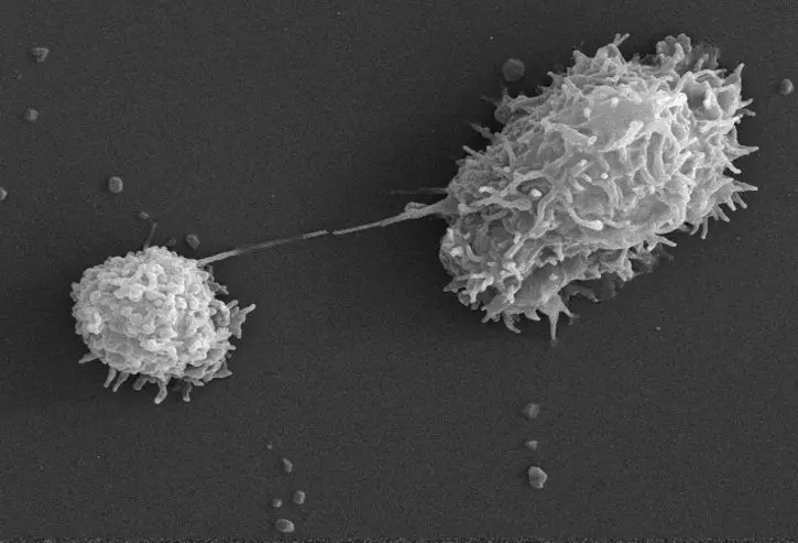
Amoeba under the microscope fixing staining techniques and structure amoeba plural amoebas amoebae is a genus that belongs to kingdom protozoa. Amoeba is an aquatic single cell unicellular organism with membrane bound eukaryotic organelles that has no definite shape. Some parasitic amoebae living inside animal bodies including humans can cause various intestinal disorders such as diarrhea ulcers and liver abscesses. Amoebas have a single cell that appears to be not much more than cytoplasm held together by a flexible cell wall. Image courtesy pearson scott foresman.
 Source: microscopemaster.com
These microscopic organisms are often called shape shifters as they have the ability to constantly change their shape. Some parasitic amoebae living inside animal bodies including humans can cause various intestinal disorders such as diarrhea ulcers and liver abscesses. Image courtesy pearson scott foresman. Stentor eats arcella youtube. The top image of amoeba proteus gives a good insight in the amoeba s anatomy.
 Source: undsci.berkeley.edu
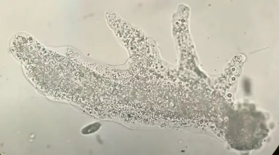
Stentor eats arcella youtube. It is a eukaryote and thus has membrane bound cell organelles and protein bound genetic material with a nuclear membrane. Image of amoeba captured with the digital ba210 microscope at 100x magnification. Amoeba under the microscope amoeba is a unicellular organism in the kingdom protozoa. Floating in this cytoplasm all kinds of cell bodies can be found.
 Source: microscopy-uk.org.uk
A tiny blob of colorless jelly with a dark speck inside it this is what an amoeba looks like when seen through a microscope. Amoebae can likewise play host to microscopic organisms that are pathogenic to people and help in spreading such microbes. When seen under a microscope the cell looks like a tiny blob of colorless jelly with a dark speck inside it. Micro 2210 study guide 2013 14 vito instructor vito at. Image of amoeba captured with the digital ba210 microscope at 100x magnification.
 Source: microscopeclarity.com
Floating in this cytoplasm all kinds of cell bodies can be found. It is capable of movement. Amoeba under the microscope fixing staining techniques structure. Onion cells and cheek cells my science blog. Amoeba under the microscope amoeba is a unicellular organism in the kingdom protozoa.
 Source: bitesizebio.com
These microscopic organisms are often called shape shifters as they have the ability to constantly change their shape. Microscopy of nosema for beekeepers. Amoeba under microscope amoeba are shapeless they look like a big blob unicellular organisms from the genus protozoa. Generally the term is used to describe single celled organisms that move in a primitive crawling manner by using temporary false feet known as pseudopods. Onion cells and cheek cells my science blog.
If you find this site value, please support us by sharing this posts to your favorite social media accounts like Facebook, Instagram and so on or you can also bookmark this blog page with the title amoeba cell under microscope by using Ctrl + D for devices a laptop with a Windows operating system or Command + D for laptops with an Apple operating system. If you use a smartphone, you can also use the drawer menu of the browser you are using. Whether it’s a Windows, Mac, iOS or Android operating system, you will still be able to bookmark this website.
