Choroid layer eye
Home » Science Education » Choroid layer eyeChoroid layer eye
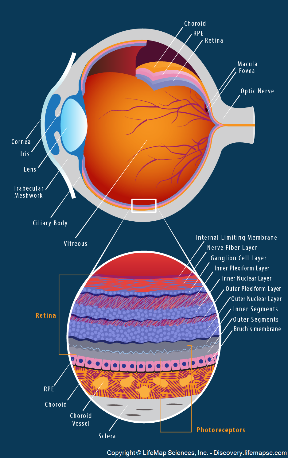
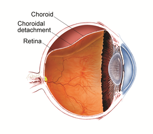
Choroid Layer Eye. It contains the retinal pigmented epithelial cells and provides oxygen and nourishment to the outer retina. The choroid is extremely vascular with its capillaries arranged in a single layer on the inner surface to nourish the outer retinal layers figure 11 14 in species with limited retinal vasculature e g horse rabbit. The uvea is the middle layer of the eye. The choroid layer also acts as a black screen which prevents extra reflections inside the eyeball so that we can get a perfect image.
 Choroidal Layers Image Layers Pandora Screenshot From pinterest.com
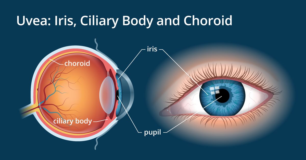
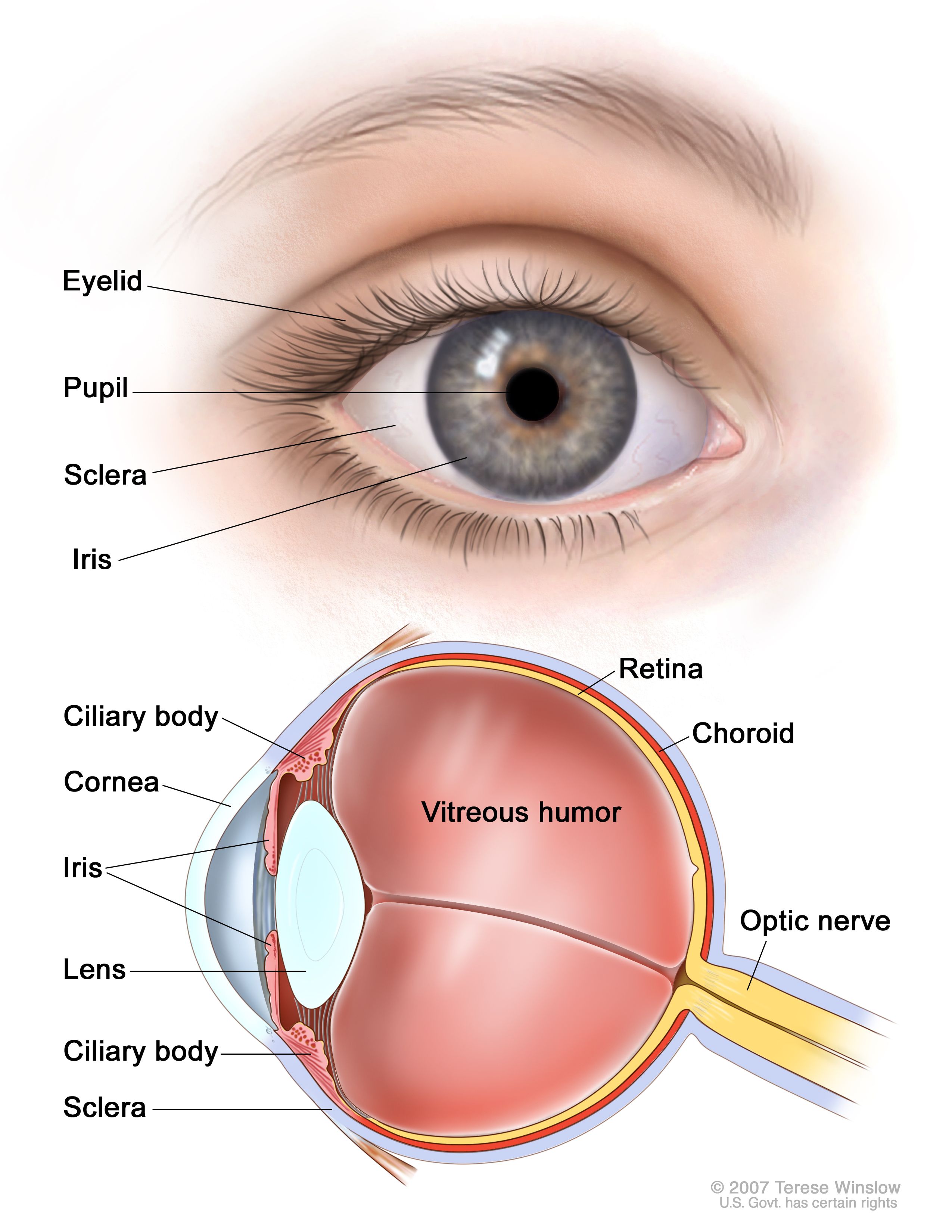
The choroid provides oxygen and nourishment to the outer layers of the retina. The uvea is the middle layer of the eye. The choroid also known as the choroidea or choroid coat is the vascular layer of the eye containing connective tissues and lying between the retina and the sclera the human choroid is thickest at the far extreme rear of the eye at 0 2 mm while in the outlying areas it narrows to 0 1 mm. It is made of the iris ciliary body and choroid. Parts are the iris ciliary body and the ligaments. Choroid is part of the uvea and supplies nutrients to the inner parts of the eye.
The choroid is extremely vascular with its capillaries arranged in a single layer on the inner surface to nourish the outer retinal layers figure 11 14 in species with limited retinal vasculature e g horse rabbit.
As a photographer i m debating this one as possibly a reasonable trade off kn. Next is an extensive capillary bed of the choriocapillary layer followed by bruch s membrane which is a common basement membrane shared by the capillary endothelial cells and the. The choroid layer also acts as a black screen which prevents extra reflections inside the eyeball so that we can get a perfect image. As a photographer i m debating this one as possibly a reasonable trade off kn. The choroid is extremely vascular with its capillaries arranged in a single layer on the inner surface to nourish the outer retinal layers figure 11 14 in species with limited retinal vasculature e g horse rabbit. This is the second layer forming the eyeball.

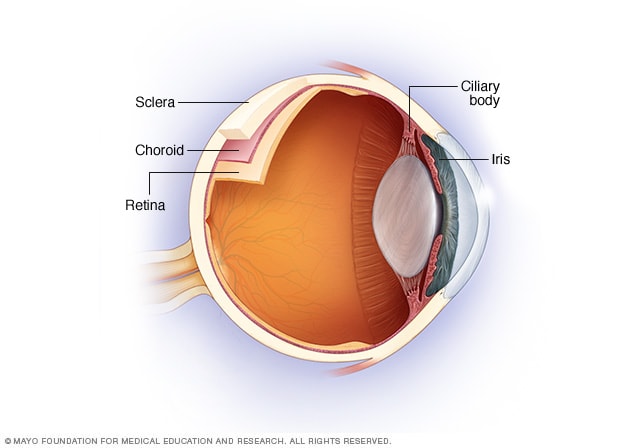
The choroid is the middle layer between sclera and retina. It is made of the iris ciliary body and choroid. It contains the retinal pigmented epithelial cells and provides oxygen and nourishment to the outer retina. The choroid also known as the choroidea or choroid coat is the vascular layer of the eye containing connective tissues and lying between the retina and the sclera the human choroid is thickest at the far extreme rear of the eye at 0 2 mm while in the outlying areas it narrows to 0 1 mm. The choroid layer also acts as a black screen which prevents extra reflections inside the eyeball so that we can get a perfect image.
 Source: discovery.lifemapsc.com
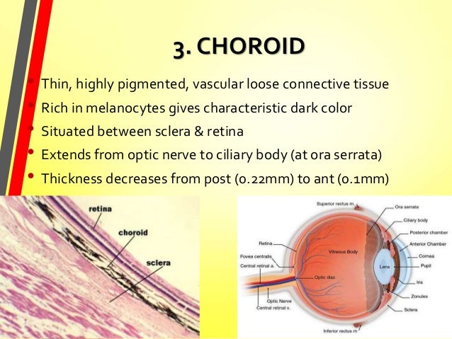
The choroid also known as the choroidea or choroid coat is the vascular layer of the eye containing connective tissues and lying between the retina and the sclera the human choroid is thickest at the far extreme rear of the eye at 0 2 mm while in the outlying areas it narrows to 0 1 mm. The choroid also known as the choroidea or choroid coat is the vascular layer of the eye containing connective tissues and lying between the retina and the sclera the human choroid is thickest at the far extreme rear of the eye at 0 2 mm while in the outlying areas it narrows to 0 1 mm. It would also mostly eliminate the retina killing it from oxygen starvation. Next is an extensive capillary bed of the choriocapillary layer followed by bruch s membrane which is a common basement membrane shared by the capillary endothelial cells and the. Closest to the connective tissue sclera is a layer of pigmented melanocytes.
 Source: en.wikipedia.org
The choroid also known as the choroidea or choroid coat is the vascular layer of the eye containing connective tissues and lying between the retina and the sclera the human choroid is thickest at the far extreme rear of the eye at 0 2 mm while in the outlying areas it narrows to 0 1 mm. It is made of the iris ciliary body and choroid. It joins the ciliary body anteriorly and lies between the retina and sclera posteriorly. It is the vascular layer of the eye containing connective tissue and it nourishes the retina and absorbs scattered light. The choroid is the middle layer between sclera and retina.
 Source: speciality.medicaldialogues.in
It is the vascular layer of the eye containing connective tissue and it nourishes the retina and absorbs scattered light. The choroid is a thin variably pigmented vascular tissue forming the posterior uvea. It joins the ciliary body anteriorly and lies between the retina and sclera posteriorly. Read an overview of general eye anatomy to learn how the parts of the eye work together. The choroid is an element of the tunica vasculosa and consists of three obvious layers.
 Source: researchgate.net
The choroid is part of the uvea and it contains blood vessels and connective tissue. It lies beneath the white part of the eye the sclera. The uvea is the middle layer of the eye. The choroid is thickest in the back of the eye where it is about 0 2 mm and narrows to 0 1 mm in the peripheral part of the eye. It consists of a densely capillary rich layer supplying blood to the eyeball.
 Source: allaboutvision.com
The choroid also known as the choroidea or choroid coat is the vascular layer of the eye containing connective tissues and lying between the retina and the sclera the human choroid is thickest at the far extreme rear of the eye at 0 2 mm while in the outlying areas it narrows to 0 1 mm. It is made of the iris ciliary body and choroid. The uvea is the middle layer of the eye. It contains the retinal pigmented epithelial cells and provides oxygen and nourishment to the outer retina. It would also mostly eliminate the retina killing it from oxygen starvation.
 Source: en.wikipedia.org
It would also mostly eliminate the retina killing it from oxygen starvation. The choroid is extremely vascular with its capillaries arranged in a single layer on the inner surface to nourish the outer retinal layers figure 11 14 in species with limited retinal vasculature e g horse rabbit. The choroid also known as the choroidea or choroid coat is the vascular layer of the eye containing connective tissues and lying between the retina and the sclera the human choroid is thickest at the far extreme rear of the eye at 0 2 mm while in the outlying areas it narrows to 0 1 mm. As a photographer i m debating this one as possibly a reasonable trade off kn. Inflammation of the choroid is called choroiditis.
 Source: eyecare-for-you.com
The choroid is thickest in the back of the eye where it is about 0 2 mm and narrows to 0 1 mm in the peripheral part of the eye. These structures control many eye functions including adjusting to different levels of light or distances of objects. The choroid provides oxygen and nourishment to the outer layers of the retina. Next is an extensive capillary bed of the choriocapillary layer followed by bruch s membrane which is a common basement membrane shared by the capillary endothelial cells and the. Choroid is part of the uvea and supplies nutrients to the inner parts of the eye.
 Source: en.wikipedia.org
The choroid is a thin variably pigmented vascular tissue forming the posterior uvea. It consists of a densely capillary rich layer supplying blood to the eyeball. The uvea is the middle layer of the eye. The choroid is an element of the tunica vasculosa and consists of three obvious layers. The choroid layer also acts as a black screen which prevents extra reflections inside the eyeball so that we can get a perfect image.
 Source: pinterest.com
Choroid is part of the uvea and supplies nutrients to the inner parts of the eye. This is the second layer forming the eyeball. The choroid provides oxygen and nourishment to the outer layers of the retina. As a photographer i m debating this one as possibly a reasonable trade off kn. The choroid is extremely vascular with its capillaries arranged in a single layer on the inner surface to nourish the outer retinal layers figure 11 14 in species with limited retinal vasculature e g horse rabbit.
 Source: asrs.org
The choroid is the middle layer between sclera and retina. Inflammation of the choroid is called choroiditis. As a photographer i m debating this one as possibly a reasonable trade off kn. The uvea is the middle layer of the eye. The choroid is the middle layer of the eye that contains blood vessels and connective tissue between the sclera the white of the eye and the retina at the back of the eye.
 Source: cancer.gov
It lies beneath the white part of the eye the sclera. It would also mostly eliminate the retina killing it from oxygen starvation. The choroid is the middle layer of the eye that contains blood vessels and connective tissue between the sclera the white of the eye and the retina at the back of the eye. It is the vascular layer of the eye containing connective tissue and it nourishes the retina and absorbs scattered light. It contains the retinal pigmented epithelial cells and provides oxygen and nourishment to the outer retina.
 Source: mayoclinic.org
Read an overview of general eye anatomy to learn how the parts of the eye work together. It lies beneath the white part of the eye the sclera. Choroid is part of the uvea and supplies nutrients to the inner parts of the eye. As a photographer i m debating this one as possibly a reasonable trade off kn. It contains the retinal pigmented epithelial cells and provides oxygen and nourishment to the outer retina.
Source: researchgate.net
It is made of the iris ciliary body and choroid. The choroid provides oxygen and nourishment to the outer layers of the retina. It would also mostly eliminate the retina killing it from oxygen starvation. The choroid is the middle layer between sclera and retina. Parts are the iris ciliary body and the ligaments.
 Source: slideshare.net
Next is an extensive capillary bed of the choriocapillary layer followed by bruch s membrane which is a common basement membrane shared by the capillary endothelial cells and the. It is made of the iris ciliary body and choroid. The choroid provides oxygen and nourishment to the outer layers of the retina. The choroid layer also acts as a black screen which prevents extra reflections inside the eyeball so that we can get a perfect image. Closest to the connective tissue sclera is a layer of pigmented melanocytes.
If you find this site convienient, please support us by sharing this posts to your preference social media accounts like Facebook, Instagram and so on or you can also save this blog page with the title choroid layer eye by using Ctrl + D for devices a laptop with a Windows operating system or Command + D for laptops with an Apple operating system. If you use a smartphone, you can also use the drawer menu of the browser you are using. Whether it’s a Windows, Mac, iOS or Android operating system, you will still be able to bookmark this website.
