Compound microscope light source
Home » Science Education » Compound microscope light sourceCompound microscope light source
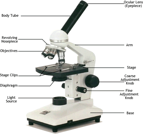
Compound Microscope Light Source. The light source is simply a light bulb. Older microscopes used mirrors to reflect light from an external source up through the bottom of the stage. A compound microscope uses a lens close to the object being viewed to collect light called the objective lens which focuses a real image of the object inside the microscope image 1. An eyepiece an additional lens is where real magnification takes place.
 The Functional Parts Of The Microscope Enfo From enfo.agt.bme.hu
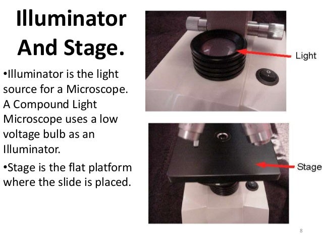
Although sometimes found as monocular with one ocular lens the compound binocular microscope is more commonly used today. An inverted microscope as the name suggests is upside down. Illuminator is the light source for a microscope typically located in the base of the microscope. Condenser is used to collect and focus the light from the illuminator on to the specimen. Older microscopes used mirrors to reflect light from an external source up through the bottom of the stage. The image of the object being viewed is enlarged because of the lens near the object.
Illuminator is the light source for a microscope typically located in the base of the microscope.
Adjusts the amount of light that reaches the specimen. Most light microscopes use low voltage halogen bulbs with continuous variable lighting control located within the base. Although sometimes found as monocular with one ocular lens the compound binocular microscope is more commonly used today. The illuminator as you can probably derive from the name is the light source of the microscope. A compound light microscope has its own light source in its base. That image is then magnified by a second lens or group of lenses called the eyepiece that gives the viewer an enlarged inverted virtual image of the object image 2.
 Source: brainly.com
Older microscopes used mirrors to reflect light from an external source up through the bottom of the stage. Illuminator is the light source for a microscope typically located in the base of the microscope. That image is then magnified by a second lens or group of lenses called the eyepiece that gives the viewer an enlarged inverted virtual image of the object image 2. The illuminator as you can probably derive from the name is the light source of the microscope. It is called a compound microscope because it compounds the light as it passes through the lenses to magnify.
 Source: microscopemaster.com
Most microscopes have a built in 110 volt steady light source that shines up through the microscope stage aperture. Illuminator is the light source for a microscope typically located in the base of the microscope. The incandescent light from the light source is reflected by a condenser lens beneath the specimen and the light passes through the specimen up to the objective lens then the projector lens sends the magnified image onto the eyepiece. Although sometimes found as monocular with one ocular lens the compound binocular microscope is more commonly used today. Adjusts the amount of light that reaches the specimen.
 Source: enfo.agt.bme.hu
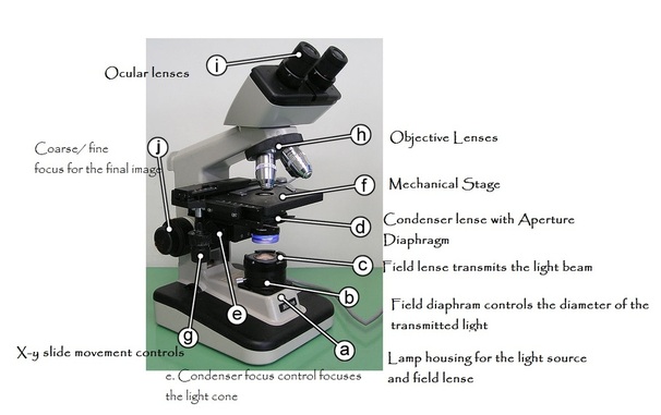
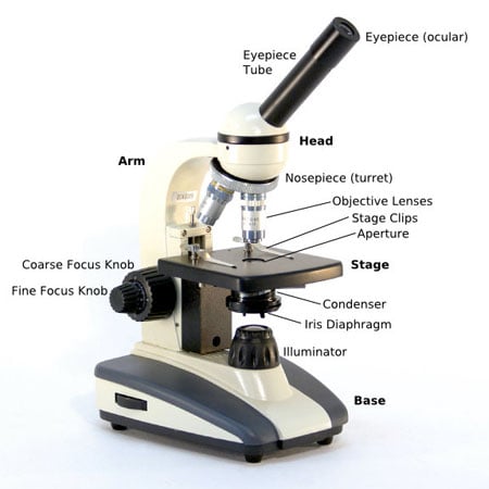
The light source iris diaphragm and condenser are the compound microscope parts responsible for illuminating the specimen. The image of the object being viewed is enlarged because of the lens near the object. Most microscopes have a built in 110 volt steady light source that shines up through the microscope stage aperture. Light from the bulb is passed through the aperture a tool that can open and close to create a beam of light of different diameters. A compound light microscope is a microscope with more than one lens and its own light source.
 Source: abacus.bates.edu
Most microscopes have a built in 110 volt steady light source that shines up through the microscope stage aperture. In this type of microscope there are ocular lenses in the binocular eyepieces and objective lenses in a rotating nosepiece closer to the specimen. Illuminator is the light source for a microscope typically located in the base of the microscope. An eyepiece an additional lens is where real magnification takes place. An inverted microscope as the name suggests is upside down.
 Source: microbiologynote.com
Compound microscope it has two convex lenses. That image is then magnified by a second lens or group of lenses called the eyepiece that gives the viewer an enlarged inverted virtual image of the object image 2. The illuminator as you can probably derive from the name is the light source of the microscope. Although sometimes found as monocular with one ocular lens the compound binocular microscope is more commonly used today. It is located under the stage often in conjunction with an iris diaphragm.
 Source: blog.microscopeworld.com
A compound microscope uses a lens close to the object being viewed to collect light called the objective lens which focuses a real image of the object inside the microscope image 1. A compound microscope can be categorized into an upright microscope and an inverted microscope. An upright microscope is just like an ordinary microscope with the lens system followed by the stage where the specimen is kept and then the light source. A compound microscope uses a lens close to the object being viewed to collect light called the objective lens which focuses a real image of the object inside the microscope image 1. A compound light microscope has its own light source in its base.
Source: math.ubc.ca
An eyepiece an additional lens is where real magnification takes place. It is located under the stage often in conjunction with an iris diaphragm. An inverted microscope as the name suggests is upside down. The light source iris diaphragm and condenser are the compound microscope parts responsible for illuminating the specimen. An eyepiece an additional lens is where real magnification takes place.
Source: insidechem.blogspot.com
However most microscopes now use a low voltage bulb. Adjusts the amount of light that reaches the specimen. The light source iris diaphragm and condenser are the compound microscope parts responsible for illuminating the specimen. Most microscopes have a built in 110 volt steady light source that shines up through the microscope stage aperture. Most light microscopes use low voltage halogen bulbs with continuous variable lighting control located within the base.
 Source: www2.nau.edu
Condenser is used to collect and focus the light from the illuminator on to the specimen. However most microscopes now use a low voltage bulb. Light from the bulb is passed through the aperture a tool that can open and close to create a beam of light of different diameters. The image of the object being viewed is enlarged because of the lens near the object. Most microscopes have a built in 110 volt steady light source that shines up through the microscope stage aperture.
 Source: microbenotes.com
The incandescent light from the light source is reflected by a condenser lens beneath the specimen and the light passes through the specimen up to the objective lens then the projector lens sends the magnified image onto the eyepiece. An eyepiece an additional lens is where real magnification takes place. In this type of microscope there are ocular lenses in the binocular eyepieces and objective lenses in a rotating nosepiece closer to the specimen. A compound light microscope has its own light source in its base. That image is then magnified by a second lens or group of lenses called the eyepiece that gives the viewer an enlarged inverted virtual image of the object image 2.
 Source: slideshare.net
Compound microscope it has two convex lenses. A compound light microscope has its own light source in its base. That image is then magnified by a second lens or group of lenses called the eyepiece that gives the viewer an enlarged inverted virtual image of the object image 2. The illuminator as you can probably derive from the name is the light source of the microscope. The light source is simply a light bulb.
 Source: fungioz.com
The light source iris diaphragm and condenser are the compound microscope parts responsible for illuminating the specimen. An eyepiece an additional lens is where real magnification takes place. The incandescent light from the light source is reflected by a condenser lens beneath the specimen and the light passes through the specimen up to the objective lens then the projector lens sends the magnified image onto the eyepiece. Light from the bulb is passed through the aperture a tool that can open and close to create a beam of light of different diameters. Illuminator is the light source for a microscope typically located in the base of the microscope.
 Source: microscope.com
Adjusts the amount of light that reaches the specimen. The illuminator as you can probably derive from the name is the light source of the microscope. Although sometimes found as monocular with one ocular lens the compound binocular microscope is more commonly used today. It is called a compound microscope because it compounds the light as it passes through the lenses to magnify. Illuminator is the light source for a microscope typically located in the base of the microscope.
 Source: microbenotes.com
Adjusts the amount of light that reaches the specimen. A compound light microscope has its own light source in its base. The light source for a microscope. Illuminator is the light source for a microscope typically located in the base of the microscope. An eyepiece an additional lens is where real magnification takes place.
 Source: slideplayer.com
However most microscopes now use a low voltage bulb. A compound light microscope has its own light source in its base. That image is then magnified by a second lens or group of lenses called the eyepiece that gives the viewer an enlarged inverted virtual image of the object image 2. A compound microscope can be categorized into an upright microscope and an inverted microscope. Adjusts the amount of light that reaches the specimen.
If you find this site good, please support us by sharing this posts to your preference social media accounts like Facebook, Instagram and so on or you can also save this blog page with the title compound microscope light source by using Ctrl + D for devices a laptop with a Windows operating system or Command + D for laptops with an Apple operating system. If you use a smartphone, you can also use the drawer menu of the browser you are using. Whether it’s a Windows, Mac, iOS or Android operating system, you will still be able to bookmark this website.
