Eyeball anatomy diagram
Home » Science Education » Eyeball anatomy diagramEyeball anatomy diagram
Eyeball Anatomy Diagram. Six extraocular muscles in the orbit are attached to the eye. Found within two cavities in the skull known as the orbits the eyes are surrounded by several supporting structures including muscles vessels and nerves. The human eye is an organ that responds to light. The orbit is formed by the cheekbone the forehead the temple and the side of the nose.
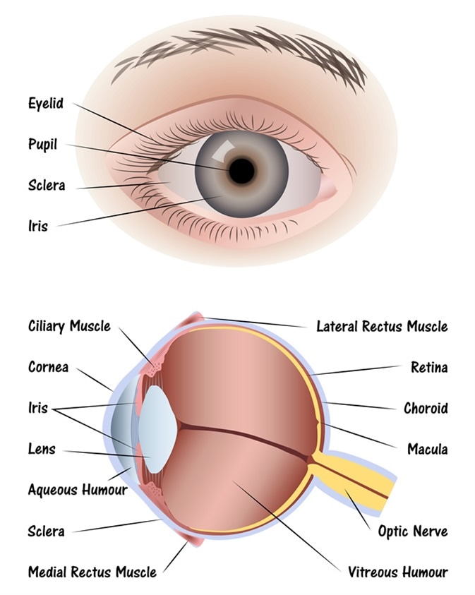 Anatomy Of The Human Eye From news-medical.net
The eyes human anatomy. In addition to the eyeball itself the orbit contains the muscles that move the eye blood vessels and nerves. It is continuous with the cornea the junction between sclera and cornea is limbus. The extraocular muscles are attached to the white part of the eye called the sclera. The human eye is an organ that responds to light. The orbit is formed by the cheekbone the forehead the temple and the side of the nose.
Basically sclera forms more than 80 of the surface area of the eyeball extending from cornea to optic nerve back of the eye.
It lies in a bony cavity within the facial skeleton or is usually known as the bony orbit. The eyes human anatomy. Wall of the eyeball. Six extraocular muscles in the orbit are attached to the eye. The eye is cushioned within the orbit by pads of fat. The eye sits in a protective bony socket called the orbit.
 Source: medicinenet.com
Basically sclera forms more than 80 of the surface area of the eyeball extending from cornea to optic nerve back of the eye. Your eyes are the windows to. In the diagram above anatomy of the eye the artery is shown in red while the vein is shown in blue. Basically sclera forms more than 80 of the surface area of the eyeball extending from cornea to optic nerve back of the eye. It is continuous with the cornea the junction between sclera and cornea is limbus.
 Source: glaucoma.org
The eyeball can be divided into three parts. What eye problems look like. Found within two cavities in the skull known as the orbits the eyes are surrounded by several supporting structures including muscles vessels and nerves. It acts as a supporting wall of the eyeball. How your eye muscles help you see.
 Source: aboutkidshealth.ca
It is the gateway to all of our five senses which allows for the perception of light awareness of colour and depth perception. This is a small tube that runs from the eye to the nasal cavity. Diagram of the different layers of the eyeball. It is the gateway to all of our five senses which allows for the perception of light awareness of colour and depth perception. Basically sclera forms more than 80 of the surface area of the eyeball extending from cornea to optic nerve back of the eye.
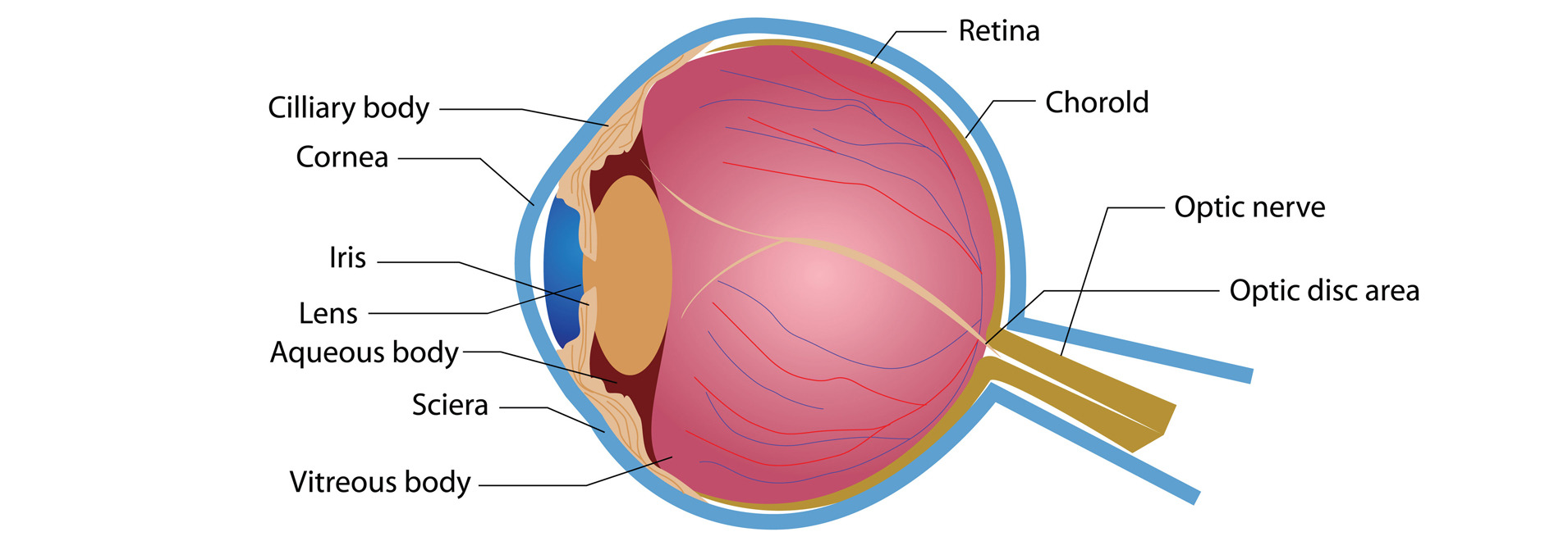 Source: eyecareproject.com
Eye fact or fiction. This is a strong layer of tissue that covers nearly the entire. Tear drains from the eyes in to the nose through the tear duct. It acts as a supporting wall of the eyeball. The orbit is formed by the cheekbone the forehead the temple and the side of the nose.
 Source: pinterest.com
The wall of the eyeball is made up of three layers fibrous outer vascular muscular middle and sensorineural inner layers. It is continuous with the cornea the junction between sclera and cornea is limbus. The human eye diagram helps in creating a schematic representation and understand the core functioning of the eye. The sclera along with the iop of the eye maintains the shape of the eyeball. This is a small tube that runs from the eye to the nasal cavity.
 Source: pinterest.com
This is why a teary eye is usually accompanied by a runny nose. The human eye is an organ that responds to light. These muscles move the eye up and down and side to side and rotate the eye. It lies in a bony cavity within the facial skeleton or is usually known as the bony orbit. It is the gateway to all of our five senses which allows for the perception of light awareness of colour and depth perception.
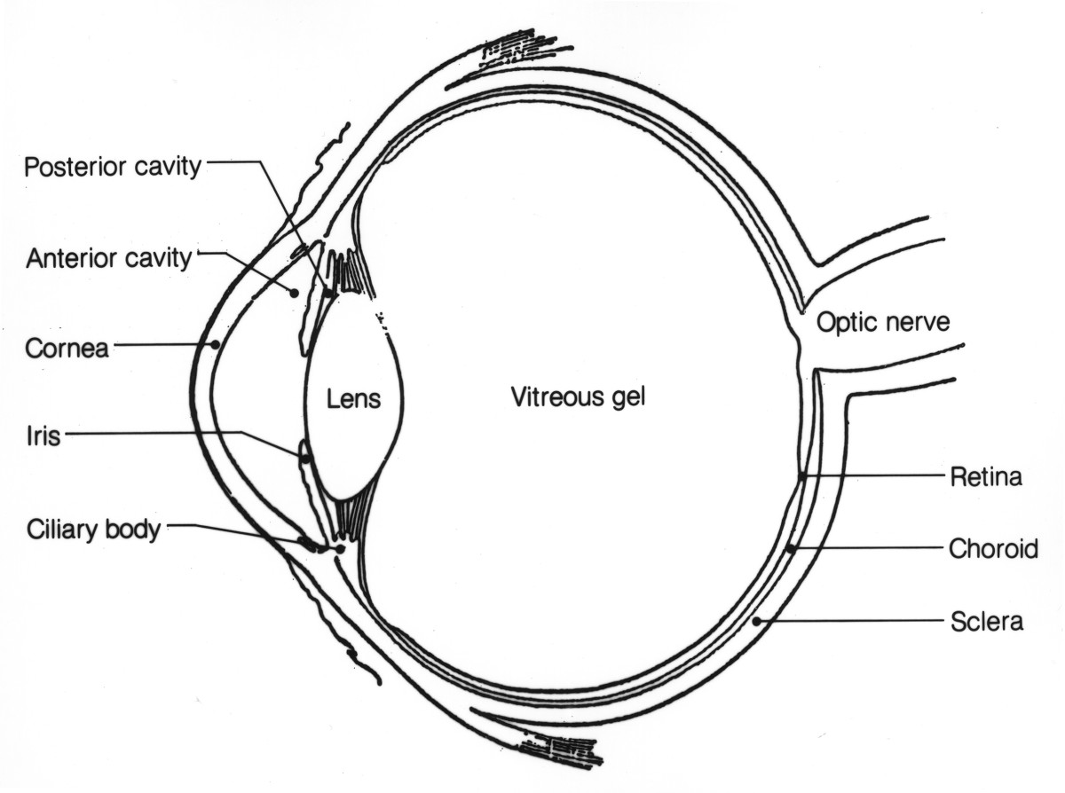 Source: owlcation.com
If you observe the eye ball diagram above the eyeball is a bilateral and spherical organ which houses the structures responsible for vision. If you observe the eye ball diagram above the eyeball is a bilateral and spherical organ which houses the structures responsible for vision. It is the gateway to all of our five senses which allows for the perception of light awareness of colour and depth perception. The eyeball has three layers each of which has several important structures that are essential for the sense of vision. The human eye is an organ that responds to light.
 Source: news-medical.net
Found within two cavities in the skull known as the orbits the eyes are surrounded by several supporting structures including muscles vessels and nerves. Diagram of the different layers of the eyeball. The fibrous vascular and inner layers. It is the gateway to all of our five senses which allows for the perception of light awareness of colour and depth perception. These muscles move the eye up and down and side to side and rotate the eye.
 Source: vision-and-eye-health.com
Wall of the eyeball. Your eyes are the windows to. The wall of the eyeball is made up of three layers fibrous outer vascular muscular middle and sensorineural inner layers. Six extraocular muscles in the orbit are attached to the eye. It is continuous with the cornea the junction between sclera and cornea is limbus.
 Source: bellboothsirkka.com
It lies in a bony cavity within the facial skeleton or is usually known as the bony orbit. How your eye muscles help you see. The human eye diagram helps in creating a schematic representation and understand the core functioning of the eye. The eye sits in a protective bony socket called the orbit. Jan 15 2013 click on various parts of our human eye illustration for descriptions of the eye anatomy.
 Source: thoughtco.com
It is the gateway to all of our five senses which allows for the perception of light awareness of colour and depth perception. Diagram function definition and eye problems. The orbit is the bony eye socket of the skull. Read an article about how vision works. In addition to the eyeball itself the orbit contains the muscles that move the eye blood vessels and nerves.
 Source: macular.org
The eye is cushioned within the orbit by pads of fat. What eye problems look like. This is why a teary eye is usually accompanied by a runny nose. The eye is cushioned within the orbit by pads of fat. The sclera along with the iop of the eye maintains the shape of the eyeball.
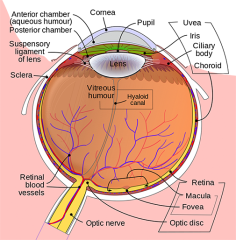 Source: umkelloggeye.org
Diagram function definition and eye problems. The eye is cushioned within the orbit by pads of fat. The eyeball can be divided into three parts. The eyes human anatomy. In the diagram above anatomy of the eye the artery is shown in red while the vein is shown in blue.
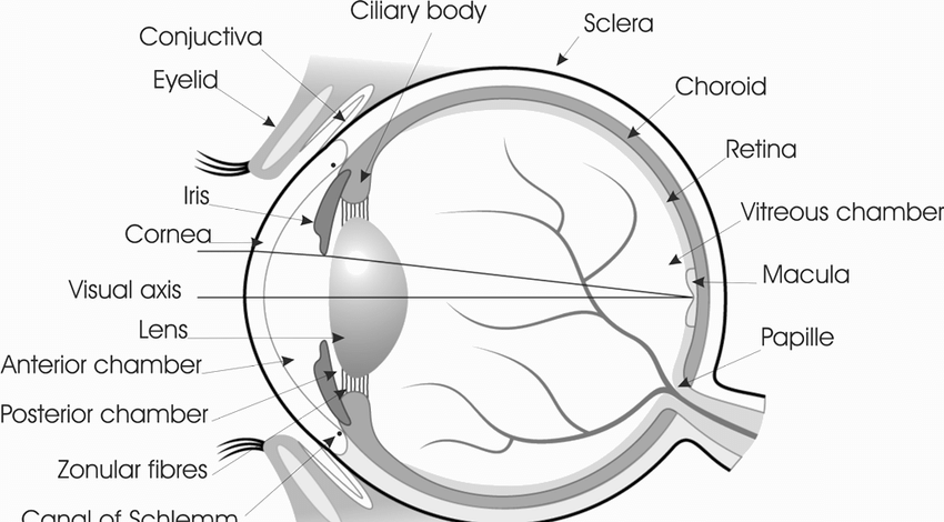 Source: lasikplus.com
Basically sclera forms more than 80 of the surface area of the eyeball extending from cornea to optic nerve back of the eye. Tear drains from the eyes in to the nose through the tear duct. The eye sits in a protective bony socket called the orbit. The orbit is the bony eye socket of the skull. In addition to the eyeball itself the orbit contains the muscles that move the eye blood vessels and nerves.
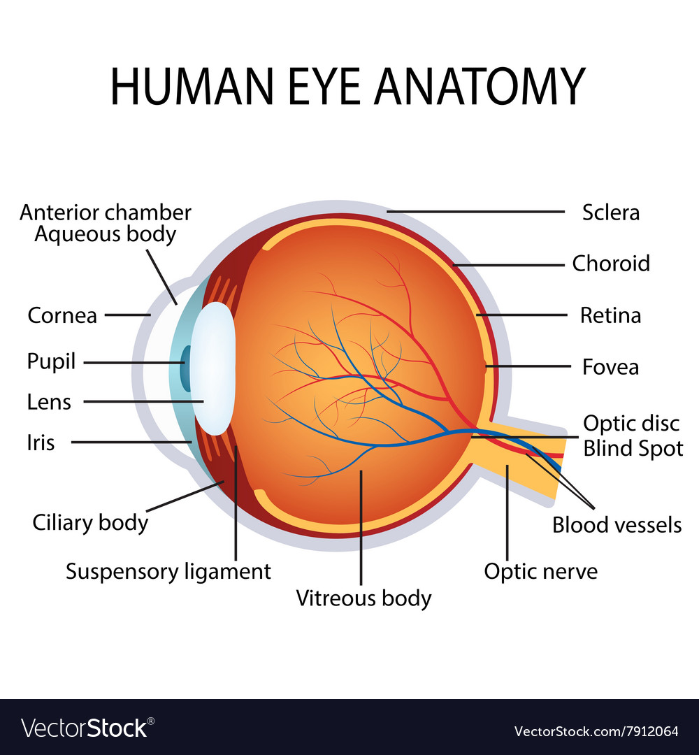 Source: vectorstock.com
Your eyes are the windows to. The fibrous vascular and inner layers. Found within two cavities in the skull known as the orbits the eyes are surrounded by several supporting structures including muscles vessels and nerves. Tear drains from the eyes in to the nose through the tear duct. It acts as a supporting wall of the eyeball.
If you find this site serviceableness, please support us by sharing this posts to your favorite social media accounts like Facebook, Instagram and so on or you can also save this blog page with the title eyeball anatomy diagram by using Ctrl + D for devices a laptop with a Windows operating system or Command + D for laptops with an Apple operating system. If you use a smartphone, you can also use the drawer menu of the browser you are using. Whether it’s a Windows, Mac, iOS or Android operating system, you will still be able to bookmark this website.
