Fetal pig dissection heart diagram
Home » Science Education » Fetal pig dissection heart diagramFetal pig dissection heart diagram
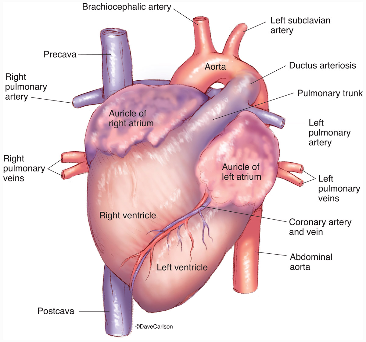
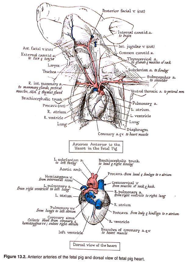
Fetal Pig Dissection Heart Diagram. Identify on your fetal pig each structure from the labeled photographs. In this activity you will open the abdominal and thoracic cavity of the fetal pig and identify structures. A pig s heart is. The heart and blood vessels of the neck region have been removed so that the trachea can be seen more clearly.
 Fetal Pig Carlson Stock Art From carlsonstockart.com
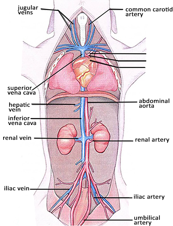
The heart and blood vessels of the neck region have been removed so that the trachea can be seen more clearly. It is opposite the dorsal side. In this activity you will open the abdominal and thoracic cavity of the fetal pig and identify structures. This diagram shows that the ductus arteriosus connects the pulmonary artery to the aorta and diverts. The pig in figure 1 is lying on its dorsal side. Be sure to follow all directions.
A pig s heart is.
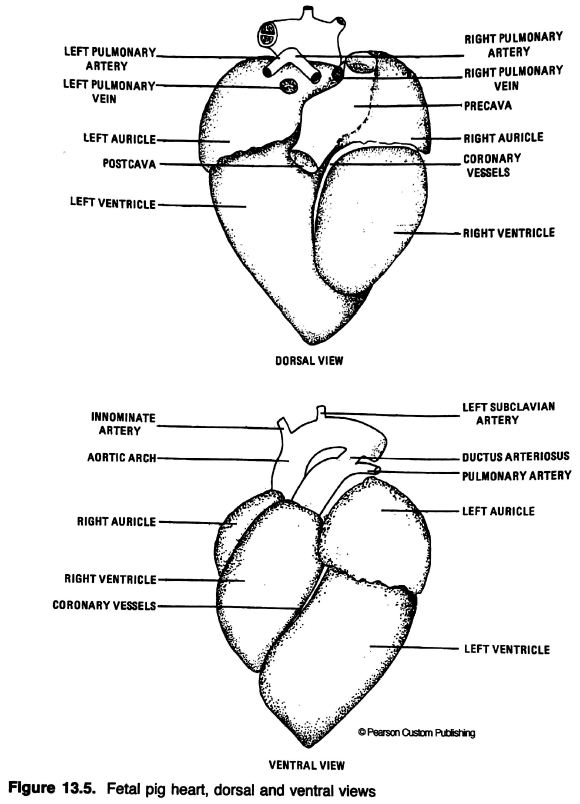
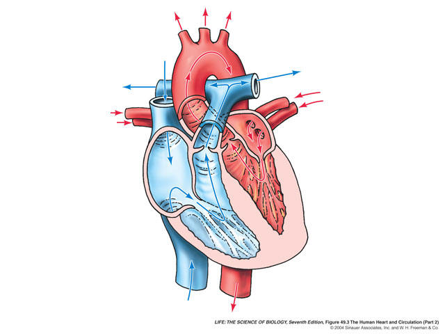
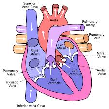
This diagram shows that the ductus arteriosus connects the pulmonary artery to the aorta and diverts. In this activity you will open the abdominal and thoracic cavity of the fetal pig and identify structures. The pig in figure 1 is lying on its dorsal side. Heart diagram fetal pig dissection pictures a pig has double system which can make blood circulate the whole body via the vessels. It is opposite the dorsal side. The aim of this experiment was to understand the external and internal structures by dissecting a pig s heart drawing and labelling the structures.
 Source: blog.valdosta.edu
Obtain a fetal pig and identify the structures listed in figure 1. The pig in figure 1 is lying on its dorsal side. The heart and blood vessels of the neck region have been removed so that the trachea can be seen more clearly. Remember that to dissect means to expose to view a careful dissection will make it easier for you to find the organs and structures. A pig s heart is.
 Source: biologycorner.com
It is opposite the dorsal side. This diagram shows that the ductus arteriosus connects the pulmonary artery to the aorta and diverts. Obtain a fetal pig and identify the structures listed in figure 1. Remember that to dissect means to expose to view a careful dissection will make it easier for you to find the organs and structures. The pig in figure 1 below has its ventral side up.
 Source: blog.valdosta.edu
Download and print the diagram for your reference. A pig s heart is. It is opposite the dorsal side. In this activity you will open the abdominal and thoracic cavity of the fetal pig and identify structures. Heart diagram fetal pig dissection pictures a pig has double system which can make blood circulate the whole body via the vessels.
 Source: pinterest.com
The pig in figure 1 below has its ventral side up. In this activity you will open the abdominal and thoracic cavity of the fetal pig and identify structures. Be sure to follow all directions. The anatomy of the fetal pig. Download and print the diagram for your reference.
 Source: quizlet.com
Download and print the diagram for your reference. The aim of this experiment was to understand the external and internal structures by dissecting a pig s heart drawing and labelling the structures. A pig s heart is. This diagram shows that the ductus arteriosus connects the pulmonary artery to the aorta and diverts. Be sure to follow all directions.

The anatomy of the fetal pig. The pig in figure 1 below has its ventral side up. Obtain a fetal pig and identify the structures listed in figure 1. The pig in figure 1 is lying on its dorsal side. With these fetal pig diagrams students are able to gain a greater understanding of the fetal pig dissection and anatomy through understanding the illustration shown in the diagram.
 Source: quizlet.com
The heart and blood vessels of the neck region have been removed so that the trachea can be seen more clearly. The aim of this experiment was to understand the external and internal structures by dissecting a pig s heart drawing and labelling the structures. Be sure to follow all directions. The pig in figure 1 is lying on its dorsal side. This diagram shows that the ductus arteriosus connects the pulmonary artery to the aorta and diverts.
 Source: courses.lumenlearning.com
Remember that to dissect means to expose to view a careful dissection will make it easier for you to find the organs and structures. The pig in figure 1 below has its ventral side up. A pig s heart is. Successfully complete dissection of the fetal pig. Remember that to dissect means to expose to view a careful dissection will make it easier for you to find the organs and structures.
 Source: courses.lumenlearning.com
Be sure to follow all directions. The aim of this experiment was to understand the external and internal structures by dissecting a pig s heart drawing and labelling the structures. Download and print the diagram for your reference. Be sure to follow all directions. The anatomy of the fetal pig.
 Source: courses.lumenlearning.com
This diagram shows that the ductus arteriosus connects the pulmonary artery to the aorta and diverts. This diagram shows that the ductus arteriosus connects the pulmonary artery to the aorta and diverts. The pig in figure 1 below has its ventral side up. Ventral is the belly side. Obtain a fetal pig and identify the structures listed in figure 1.
 Source: courses.lumenlearning.com
It is opposite the dorsal side. Obtain a fetal pig and identify the structures listed in figure 1. The aim of this experiment was to understand the external and internal structures by dissecting a pig s heart drawing and labelling the structures. The pig in figure 1 is lying on its dorsal side. Use figures 1 4 below to identify its sex.
 Source: carlsonstockart.com
With these fetal pig diagrams students are able to gain a greater understanding of the fetal pig dissection and anatomy through understanding the illustration shown in the diagram. In this activity you will open the abdominal and thoracic cavity of the fetal pig and identify structures. Heart diagram fetal pig dissection pictures a pig has double system which can make blood circulate the whole body via the vessels. The anatomy of the fetal pig. Identify on your fetal pig each structure from the labeled photographs.
 Source: people.hsc.edu
Use figures 1 4 below to identify its sex. The heart and blood vessels of the neck region have been removed so that the trachea can be seen more clearly. Ventral is the belly side. The pig in figure 1 is lying on its dorsal side. Identify on your fetal pig each structure from the labeled photographs.
 Source: journeythroughbiology.wordpress.com
With these fetal pig diagrams students are able to gain a greater understanding of the fetal pig dissection and anatomy through understanding the illustration shown in the diagram. This diagram shows that the ductus arteriosus connects the pulmonary artery to the aorta and diverts. In this activity you will open the abdominal and thoracic cavity of the fetal pig and identify structures. The pig in figure 1 below has its ventral side up. It is opposite the dorsal side.
 Source: pinterest.com
The pig in figure 1 is lying on its dorsal side. The pig in figure 1 below has its ventral side up. Identify on your fetal pig each structure from the labeled photographs. This diagram shows that the ductus arteriosus connects the pulmonary artery to the aorta and diverts. The heart and blood vessels of the neck region have been removed so that the trachea can be seen more clearly.
If you find this site value, please support us by sharing this posts to your favorite social media accounts like Facebook, Instagram and so on or you can also save this blog page with the title fetal pig dissection heart diagram by using Ctrl + D for devices a laptop with a Windows operating system or Command + D for laptops with an Apple operating system. If you use a smartphone, you can also use the drawer menu of the browser you are using. Whether it’s a Windows, Mac, iOS or Android operating system, you will still be able to bookmark this website.
