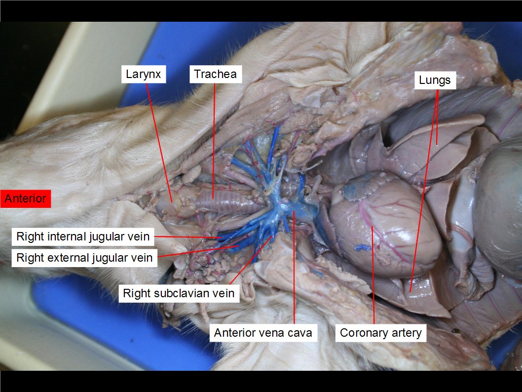
Fetal pig lungs
Home » Science Education » Fetal pig lungsFetal pig lungs
Fetal Pig Lungs. The parietal pleura is a serous membrane which surrounds the lungs is shown being lifted up by the straight teasing needle it is like a thin film and can be somewhat difficult to remove and isolate. How many chambers does the pig heart have. The visceral pleura is seen on the layer underneath. The lungs have the responsibilty of removing carbon dioxide and adding oxygen to blood that will then be distributed back to the body through the capillaries.
 Reading Fetal Pig Dissection Biology Ii Laboratory Manual From courses.lumenlearning.com
The pig in the first photograph below has its ventral side up. The bronchi branch further inside the lungs becoming bronchioles which terminate at alveoli clusters of air sacs where oxygen and carbon dioxide are exchanged with tiny. It is opposite the dorsal side. Focus next on the abdominal cavity. Digestive system urinary system. What cavity contains the lungs.
Obtain a fetal pig and identify the structures listed in figure 1.
The fetal pigs lungs are flatter than those of a human. The lungs are found in the thoracic cavity and can be accessed by breaking the thin layer of connective tissue known as the pleura. First look at the digestive system organs. How does the size of the pig lungs compare to the size of the frog lungs you dissected previously. Use the photographs below to identify its sex. Air from the oral and nasal passages enters the lungs via the trachea which branches into two bronchi as it enters the lungs.
 Source: youtube.com
Fetal pig being dissected showing the thoracic cavity including the rib cage the heart in the pericardium the lungs the diaphragm part of the liver and all the organs under the neck. Air from the oral and nasal passages enters the lungs via the trachea which branches into two bronchi as it enters the lungs. Techniques are being mastered to transplant porcine pig lungs into humans. This picture shows a dorsal view of the respiratory organs of the pig removed from the thoracic cavity. The aortic arch and the aorta are to the left of the trachea and the aorta has been cut.
 Source: courses.lumenlearning.com
How many chambers does the pig heart have. The tachea is the windpipe that inhaled air passes through to get to the lungs. Follow the trachea to where it branches into two bronchi and observe that each bronchus leads to a lung. Fetal pig being dissected showing the thoracic cavity including the rib cage the heart in the pericardium the lungs the diaphragm part of the liver and all the organs under the neck. Focus next on the abdominal cavity.
 Source: courses.lumenlearning.com
The pig in the first photograph below has its ventral side up. The parietal pleura is shown on the thoracic cavity wall in this picture because it was attached to the wall and ripped from the lung surface. The visceral pleura is seen on the layer underneath. The pig in the first photograph below is laying on its dorsal side. The lungs are found in the thoracic cavity and can be accessed by breaking the thin layer of connective tissue known as the pleura.
 Source: chem.libretexts.org
What cavity contains the lungs. It is ringed and rigid and is called the windpipe in contrast to the esophagus which is composed of smooth muscle. The pig in the first photograph below has its ventral side up. Each lung is located in a body cavity called a pleural cavity. It runs from the larynx to the lungs.
 Source: schoolworkhelper.net
How does the size of the pig lungs compare to the size of the frog lungs you dissected previously. Techniques are being mastered to transplant porcine pig lungs into humans. Digestive system urinary system. Use the photographs below to identify its sex. Dissection of a fetal pig.

It is opposite the dorsal side. The parietal pleura is shown on the thoracic cavity wall in this picture because it was attached to the wall and ripped from the lung surface. How does the size of the pig lungs compare to the size of the frog lungs you dissected previously. Fetal pig being dissected showing the thoracic cavity including the rib cage the heart in the pericardium the lungs the diaphragm part of the liver and all the organs under the neck. The lungs have the responsibilty of removing carbon dioxide and adding oxygen to blood that will then be distributed back to the body through the capillaries.

The visceral pleura is seen on the layer underneath. Dissection of a fetal pig. The bronchi branch further inside the lungs becoming bronchioles which terminate at alveoli clusters of air sacs where oxygen and carbon dioxide are exchanged with tiny. Respiratory system lungs. Follow the trachea to where it branches into two bronchi and observe that each bronchus leads to a lung.

How many chambers does the pig heart have. The size of the fetal pig depends on the time allowed for the mother to gestate. What role does the diaphragm play in respiration. It is opposite the dorsal side. The pig in the first photograph below is laying on its dorsal side.
 Source: cals.ncsu.edu
The heart is on the opposite ventral side. Fetal pig being dissected showing the thoracic cavity including the rib cage the heart in the pericardium the lungs the diaphragm part of the liver and all the organs under the neck. The aortic arch and the aorta are to the left of the trachea and the aorta has been cut. How does the size of the pig lungs compare to the size of the frog lungs you dissected previously. What cavity contains the heart.
 Source: courses.lumenlearning.com
The lungs have the responsibilty of removing carbon dioxide and adding oxygen to blood that will then be distributed back to the body through the capillaries. Digestive system urinary system. Ventral is the belly side. The fetal pigs lungs are flatter than those of a human. Obtain a fetal pig and identify the structures listed in the first photograph.
 Source: courses.lumenlearning.com
Focus next on the abdominal cavity. The visceral pleura is seen on the layer underneath. This picture shows a dorsal view of the respiratory organs of the pig removed from the thoracic cavity. The size of the fetal pig depends on the time allowed for the mother to gestate. The pig in the first photograph below is laying on its dorsal side.
 Source: courses.lumenlearning.com
The visceral pleura is seen on the layer underneath. The tachea is the windpipe that inhaled air passes through to get to the lungs. The lungs are found in the thoracic cavity and can be accessed by breaking. Respiratory system lungs. Techniques are being mastered to transplant porcine pig lungs into humans.
 Source: courses.lumenlearning.com
Respiratory system lungs. The pig in the first photograph below has its ventral side up. The lungs are found in the thoracic cavity and can be accessed by breaking. Air from the oral and nasal passages enters the lungs via the trachea which branches into two bronchi as it enters the lungs. Each lung is located in a body cavity called a pleural cavity.
 Source: courses.lumenlearning.com
The lungs are found in the thoracic cavity and can be accessed by breaking the thin layer of connective tissue known as the pleura. The trachea has been cut in half along its long axis so that its interior surface cartilaginous rings and bifurcation into the two bronchi are visible. Respiratory system lungs. Follow the trachea to where it branches into two bronchi and observe that each bronchus leads to a lung. Dissection of a fetal pig.
 Source: flickriver.com
The parietal pleura is shown on the thoracic cavity wall in this picture because it was attached to the wall and ripped from the lung surface. The lungs are found in the thoracic cavity and can be accessed by breaking the thin layer of connective tissue known as the pleura. Dissection of a fetal pig. How does the size of the pig lungs compare to the size of the frog lungs you dissected previously. What role does the diaphragm play in respiration.
If you find this site helpful, please support us by sharing this posts to your favorite social media accounts like Facebook, Instagram and so on or you can also save this blog page with the title fetal pig lungs by using Ctrl + D for devices a laptop with a Windows operating system or Command + D for laptops with an Apple operating system. If you use a smartphone, you can also use the drawer menu of the browser you are using. Whether it’s a Windows, Mac, iOS or Android operating system, you will still be able to bookmark this website.
