Fetal pig mouth
Home » Science Education » Fetal pig mouthFetal pig mouth
Fetal Pig Mouth. Compare the diagram of the fetal pig digestive system on the previous page to. Learn vocabulary terms and more with flashcards games and other study tools. The dissection of the fetal pig in the laboratory is important because pigs and. After completing the cuts locate the umbilical vein that leads from the umbilical cord to the liver.

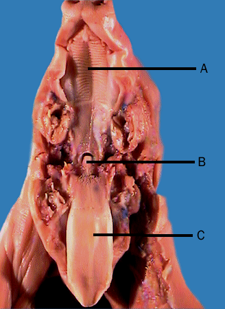
Use scissors to cut through the skin and muscles according to the diagram. Respiratory 1 mouth pharynx thorax. Obtain a fetal pig and identify the structures listed in the first photograph. The pig in the first photograph below is laying on its dorsal side. The pig in the first photograph below has its ventral side up. Open mouth of fetal pig 11.
Do not remove the umbilical cord.
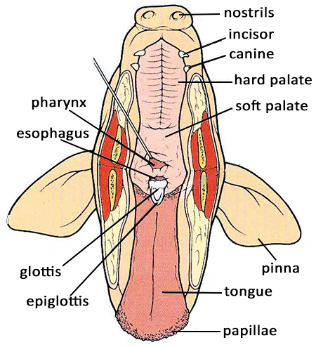
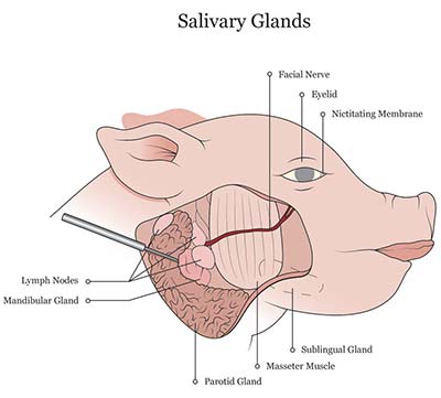
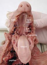
It is opposite the dorsal side. Do not remove the umbilical cord. Pay attention to the amount and color of hair birth marks and other unique markings. The hard palate makes up the anterior part of the roof of the mouth. Place your fetal pig in the dissecting pan ventral side up. The pig in the first photograph below has its ventral side up.
 Source: biologycorner.com
Open mouth of fetal pig 11. The pig in the first photograph below is laying on its dorsal side. Mammary glands later develop only in maturing females. Use the photographs below to identify its sex. Pay attention to the amount and color of hair birth marks and other unique markings.

Start studying fetal pig mouth and pharynx. Do not remove the umbilical cord. The pig in the first photograph below has its ventral side up. Respiratory 1 mouth pharynx thorax. Compare the diagram of the fetal pig digestive system on the previous page to.

Obtain a fetal pig and identify the structures listed in the first photograph. It is opposite the dorsal side. Use the photographs below to identify its sex. The esophagus carries food liquids and saliva from the mouth to the stomach. Swine are omnivores and there is probably no other animal that is quite as focused on food.

Fetal pig lab one. The pig in the first photograph below has its ventral side up. Use scissors to cut through the skin and muscles according to the diagram. You will need to cut this vein in order to open up the abdominal cavity. Ventral is the belly side.

Fetal pig lab one. After completing the cuts locate the umbilical vein that leads from the umbilical cord to the liver. Learn vocabulary terms and more with flashcards games and other study tools. Compare the diagram of the fetal pig digestive system on the previous page to. Fetal pig dissection guide.
 Source: quizlet.com
Use the photographs below to identify its sex. It is opposite the dorsal side. Respiratory 1 mouth pharynx thorax. Place your fetal pig in the dissecting pan ventral side up. The esophagus carries food liquids and saliva from the mouth to the stomach.

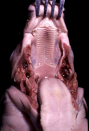
Fetal pig dissection guide. Made of bone and covered with folds of mucus membrane the hard palate separates the oral cavity from the nasal cavities. Use the photographs below to identify its sex. The pig in the first photograph below is laying on its dorsal side. Be sure to fetal pig lab one.
 Source: biologycorner.com
Respiratory 1 mouth pharynx thorax external anatomy examine the fetal pig and locate the external features shown above. Ventral is the belly side. It is opposite the dorsal side. The larynx is located between the pharynx and the trachea and holds the vocal cords. Give a pig a treat like the animal cracker in the mouth of the little pig to the right and they ll do almost anything.
 Source: quizlet.com
You will need to cut this vein in order to open up the abdominal cavity. Learn vocabulary terms and more with flashcards games and other study tools. Give a pig a treat like the animal cracker in the mouth of the little pig to the right and they ll do almost anything. Place your fetal pig in the dissecting pan ventral side up. Piglets are born with needle teeth which are the deciduous third incisors and the canines.
 Source: apbiovet.wordpress.com
Do not remove the umbilical cord. The pig in the first photograph below is laying on its dorsal side. Compare the diagram of the fetal pig digestive system on the previous page to. Dental anatomy of pigs. Studied this semester in the context of a single specimen the fetal pig.
 Source: biologycorner.com
Be sure to fetal pig lab one. Respiratory 1 mouth pharynx thorax. Start a free trial of quizlet plus by thanksgiving lock in 50 off all year try it free. Made of bone and covered with folds of mucus membrane the hard palate separates the oral cavity from the nasal cavities. Compare the diagram of the fetal pig digestive system on the previous page to.
 Source: cram.com
Start a free trial of quizlet plus by thanksgiving lock in 50 off all year try it free. Give a pig a treat like the animal cracker in the mouth of the little pig to the right and they ll do almost anything. It is opposite the dorsal side. Place your fetal pig in the dissecting pan ventral side up. External fetal pig dissection worksheet.
 Source: quizlet.com
The dissection of the fetal pig in the laboratory is important because pigs and. Start a free trial of quizlet plus by thanksgiving lock in 50 off all year try it free. Respiratory 1 mouth pharynx thorax external anatomy examine the fetal pig and locate the external features shown above. Carefully examine the external features of your pig beginning with the head. The pig in the first photograph below has its ventral side up.
 Source: learning-center.homesciencetools.com
Start a free trial of quizlet plus by thanksgiving lock in 50 off all year try it free. Fetal pig lab one. After completing the cuts locate the umbilical vein that leads from the umbilical cord to the liver. Start studying fetal pig mouth and pharynx. Studied this semester in the context of a single specimen the fetal pig.
 Source: whitman.edu
Respiratory 1 mouth pharynx thorax external anatomy examine the fetal pig and locate the external features shown above. Two rows of nipples of mammary glands are present on the ventral abdominal surface of both males and females. The hard palate makes up the anterior part of the roof of the mouth. The pig in the first photograph below is laying on its dorsal side. Carefully examine the external features of your pig beginning with the head.
If you find this site adventageous, please support us by sharing this posts to your own social media accounts like Facebook, Instagram and so on or you can also save this blog page with the title fetal pig mouth by using Ctrl + D for devices a laptop with a Windows operating system or Command + D for laptops with an Apple operating system. If you use a smartphone, you can also use the drawer menu of the browser you are using. Whether it’s a Windows, Mac, iOS or Android operating system, you will still be able to bookmark this website.
