Frog dissection manual
Home » Science Education » Frog dissection manualFrog dissection manual
Frog Dissection Manual. When done you should be able to easily open the mouth to examine these. Locate the frog s eyes the nictitating membrane is a clear membrane that is attached to the bottom of the eye. Place the frog on the dissecting pan. Use the scissors to cut the back of the mouth where the mandible attaches to the maxilla.
 Pin By Bon Heitz On Random Frog Dissection Frog Dissection Worksheet Eye Anatomy Diagram From pinterest.com
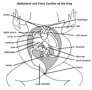
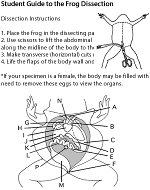
Pithing greatly reduces the incidence and intensity of muscle contractions during dissection. Head the anterior end of the frog extending to and including the eardrums. Internal anatomy frog dissection manual. Use these descriptions and the picture to identify each of the following external structures. The frog will twitch. To locate observe and diagram the external outside structures of frogs.
Use tweezers to carefully remove the nictitating membrane.
Pithing greatly reduces the incidence and intensity of muscle contractions during dissection. First weigh the frog and measure its length from snout to vent. Dorsal the back of an organism ventral the belly of an organism lateral the sides of an organism. External anatomy frog dissection manual. The frog will twitch. To see similarities between this organism and ourselves.
 Source: pinterest.com
Head the anterior end of the frog extending to and including the eardrums. The dissection begins with an anesthetized frog. Head the anterior end of the frog extending to and including the eardrums. Place the frog on the dissecting pan. Use tweezers to carefully remove the nictitating membrane.
 Source: biologyjunction.com
To locate observe and diagram the external outside structures of frogs. When done you should be able to easily open the mouth to examine these. Dorsal the back of an organism ventral the belly of an organism lateral the sides of an organism. Head the anterior end of the frog extending to and including the eardrums. Frog dissection manual.
 Source: biologycorner.com
When done you should be able to easily open the mouth to examine these. Place the frog on the dissecting pan. Head the anterior end of the frog extending to and including the eardrums. To see similarities between this organism and ourselves. Dorsal the back of an organism ventral the belly of an organism lateral the sides of an organism.
 Source: zbook.org
Compare the length of your frog to other frogs br your frog cm br frog 2 br frog 3 br frog 4 br frog 5 br average br l e n g th br 5. Pithing greatly reduces the incidence and intensity of muscle contractions during dissection. Read reviews from world s largest community for readers. Frog dissection manual book. Use these descriptions and the picture to identify each of the following external structures.

Read reviews from world s largest community for readers. Pithing greatly reduces the incidence and intensity of muscle contractions during dissection. The dissection begins with an anesthetized frog. When done you should be able to easily open the mouth to examine these. Place the frog on the dissecting pan.
 Source: pinterest.com
Dorsal the back of an organism ventral the belly of an organism lateral the sides of an organism. Read reviews from world s largest community for readers. External anatomy frog dissection manual. Compare the length of your frog to other frogs br your frog cm br frog 2 br frog 3 br frog 4 br frog 5 br average br l e n g th br 5. To see similarities between this organism and ourselves.
 Source: merospark.com
To see similarities between this organism and ourselves. To locate observe and diagram the external outside structures of frogs. Use these descriptions and the picture to identify each of the following external structures. Open the mouth using your fingers or forceps. Compare the length of your frog to other frogs br your frog cm br frog 2 br frog 3 br frog 4 br frog 5 br average br l e n g th br 5.

The dissection begins with an anesthetized frog. Do not pin it down. Internal anatomy frog dissection manual. The dissection begins with an anesthetized frog. Head the anterior end of the frog extending to and including the eardrums.
 Source: slideshare.net
Source: slideshare.net
Compare the length of your frog to other frogs br your frog cm br frog 2 br frog 3 br frog 4 br frog 5 br average br l e n g th br 5. Dorsal the back of an organism ventral the belly of an organism lateral the sides of an organism. Do not pin it down. Frog dissection manual book. Read reviews from world s largest community for readers.
 Source: yumpu.com
The frog dissection manual page 1. The dissection begins with an anesthetized frog. Place the frog on the dissecting pan. Decapitate the frog with scissors and pith the spinal cord with a pithing needle. First weigh the frog and measure its length from snout to vent.
 Source: norecopa.no
Do not pin it down. Pithing greatly reduces the incidence and intensity of muscle contractions during dissection. Internal anatomy frog dissection manual. The dissection begins with an anesthetized frog. Open the mouth using your fingers or forceps.
 Source: carolina.com
The dissection begins with an anesthetized frog. Compare the length of your frog to other frogs br your frog cm br frog 2 br frog 3 br frog 4 br frog 5 br average br l e n g th br 5. Use the scissors to cut the back of the mouth where the mandible attaches to the maxilla. Open the mouth using your fingers or forceps. Use these descriptions and the picture to identify each of the following external structures.
 Source: diagramcliffv.facieurope.it
Decapitate the frog with scissors and pith the spinal cord with a pithing needle. Frog dissection manual book. First weigh the frog and measure its length from snout to vent. Head the anterior end of the frog extending to and including the eardrums. To see similarities between this organism and ourselves.
 Source: carolina.com
When done you should be able to easily open the mouth to examine these. Do not pin it down. Use tweezers to carefully remove the nictitating membrane. Place the frog on the dissecting pan. Locate the frog s eyes the nictitating membrane is a clear membrane that is attached to the bottom of the eye.
 Source: pinterest.com
Decapitate the frog with scissors and pith the spinal cord with a pithing needle. Head the anterior end of the frog extending to and including the eardrums. When done you should be able to easily open the mouth to examine these. Use these descriptions and the picture to identify each of the following external structures. Structures inside the mouth.
If you find this site adventageous, please support us by sharing this posts to your own social media accounts like Facebook, Instagram and so on or you can also save this blog page with the title frog dissection manual by using Ctrl + D for devices a laptop with a Windows operating system or Command + D for laptops with an Apple operating system. If you use a smartphone, you can also use the drawer menu of the browser you are using. Whether it’s a Windows, Mac, iOS or Android operating system, you will still be able to bookmark this website.