Human blood smear slide
Home » Science Education » Human blood smear slideHuman blood smear slide
Human Blood Smear Slide. C m ac m a blood smear preparation materials and supplies sample edta slides fixative buffer and stain diff quik water 4. C m ac m a a spreader slide is used to feather the drop of blood. Light micrograph of a smear of human blood. Blood smear human 5.
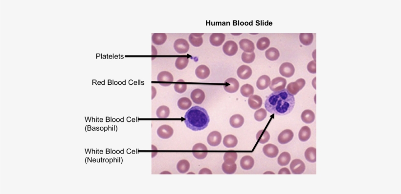 Top Images For Blood Smear Labeled On Picsunday Human Blood Slide Labeled Free Transparent Png Download Pngkey From pngkey.com
Cells will be heaped and piled close to the point were the drop of blood was placed on the slide. C m ac m a a spreader slide is used to feather the drop of blood. A poor slide is a torment. Place a small drop of blood or one side about 1 2 cm from one end. The slide from which this image was prepared was a blood smear it was made by putting a drop of blood on one end of a slide and using a second slide to spread the blood into a thin uniform layer over the slide. Eosinophil basophil click on the any of the small images above to see a higher resolution image.
3 packed in a plastic box human blood smear used to observe the cell composition of blood.
2 1 x 3 25 mm x 76 mm glass slides. Place a small drop of blood or one side about 1 2 cm from one end. 2 1 x 3 25 mm x 76 mm glass slides. Blood within 1 hr. The slide should be clean. A poor slide is a torment.
 Source: eugraph.com
The extra time and care taken during the field season will be rewarded later when the smears must be scanned and parasites identified and counted. Of collection preparation of blood film. Without delay place a spreader at an angle of 45 from the slide and move it back to make contact with the drop. Preparation of blood smear. Making and staining a blood smear a well made blood smear is a beauty to behold and likely to yield interesting and significant information for a research project.
 Source: pngkey.com
Eosinophil basophil click on the any of the small images above to see a higher resolution image. 3 packed in a plastic box human blood smear used to observe the cell composition of blood. It consists of a fluid called plasma and cells formed elements that are suspended in the plasma. Blood smear human wright s stain h6455 microscope at 1000x these images show erythrocytes red blood cells leukocytes white blood cells and platelets as they appear in a wright s stained blood smear. Of collection preparation of blood film.
 Source: fishersci.com
C m ac m a a drop of blood is placed on a clean microscope slide. Place a small drop of blood or one side about 1 2 cm from one end. Light micrograph of a smear of human blood. C m ac m a a drop of blood is placed on a clean microscope slide. White blood cells appear shrunken and some types are difficult to distinguish from each other.
 Source: fishersci.com
Place a small drop of blood or one side about 1 2 cm from one end. 3 packed in a plastic box human blood smear used to observe the cell composition of blood. Blood smear erythrocytes red blood cells 100 x. Human blood smear wrights basophil 3. Finger prick or.
 Source: amazon.com
The slide from which this image was prepared was a blood smear it was made by putting a drop of blood on one end of a slide and using a second slide to spread the blood into a thin uniform layer over the slide. Blood smear human 5. Blood within 1 hr. Place a small drop of blood or one side about 1 2 cm from one end. Other of biology slide human blood smear slide chlamydomonas slide volvox slide and liver fluke slide offered by the national scientific suppliers chennai tamil nadu.
 Source: austincc.edu
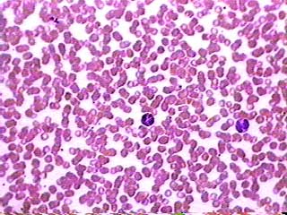
Leukocyte cell types white blood cells platlets are visible rbc. Of collection preparation of blood film. Eosinophil basophil click on the any of the small images above to see a higher resolution image. White blood cells appear shrunken and some types are difficult to distinguish from each other. 2 1 x 3 25 mm x 76 mm glass slides.
 Source: homesciencetools.com
Blood is an unusual connective tissue because it is normally in liquid form. Eosinophil basophil click on the any of the small images above to see a higher resolution image. The slide from which this image was prepared was a blood smear it was made by putting a drop of blood on one end of a slide and using a second slide to spread the blood into a thin uniform layer over the slide. Blood smear human 5. The slide should be clean.
 Source: austincc.edu
Where to look for cells in a blood smear the density of cells varies across the smear. Place a small drop of blood or one side about 1 2 cm from one end. Cells will be heaped and piled close to the point were the drop of blood was placed on the slide. Blood smear human 5. Blood within 1 hr.
 Source: labtestsonline.org
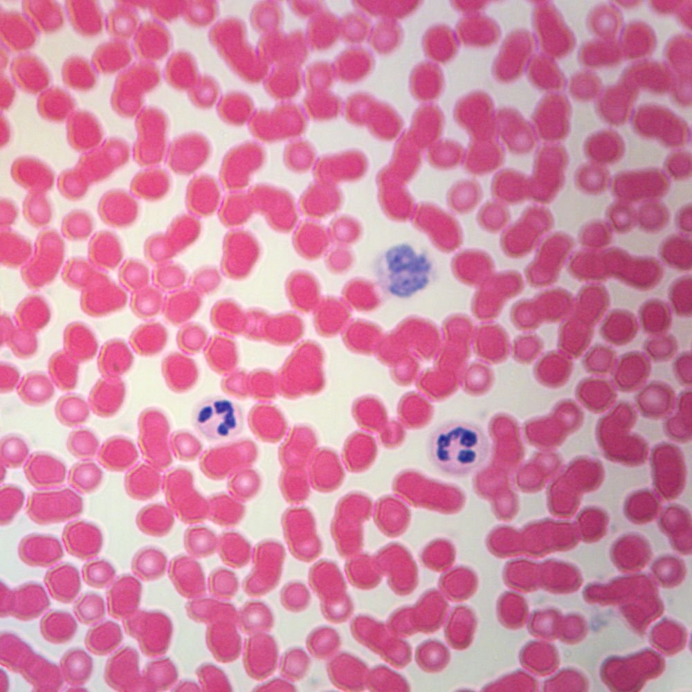
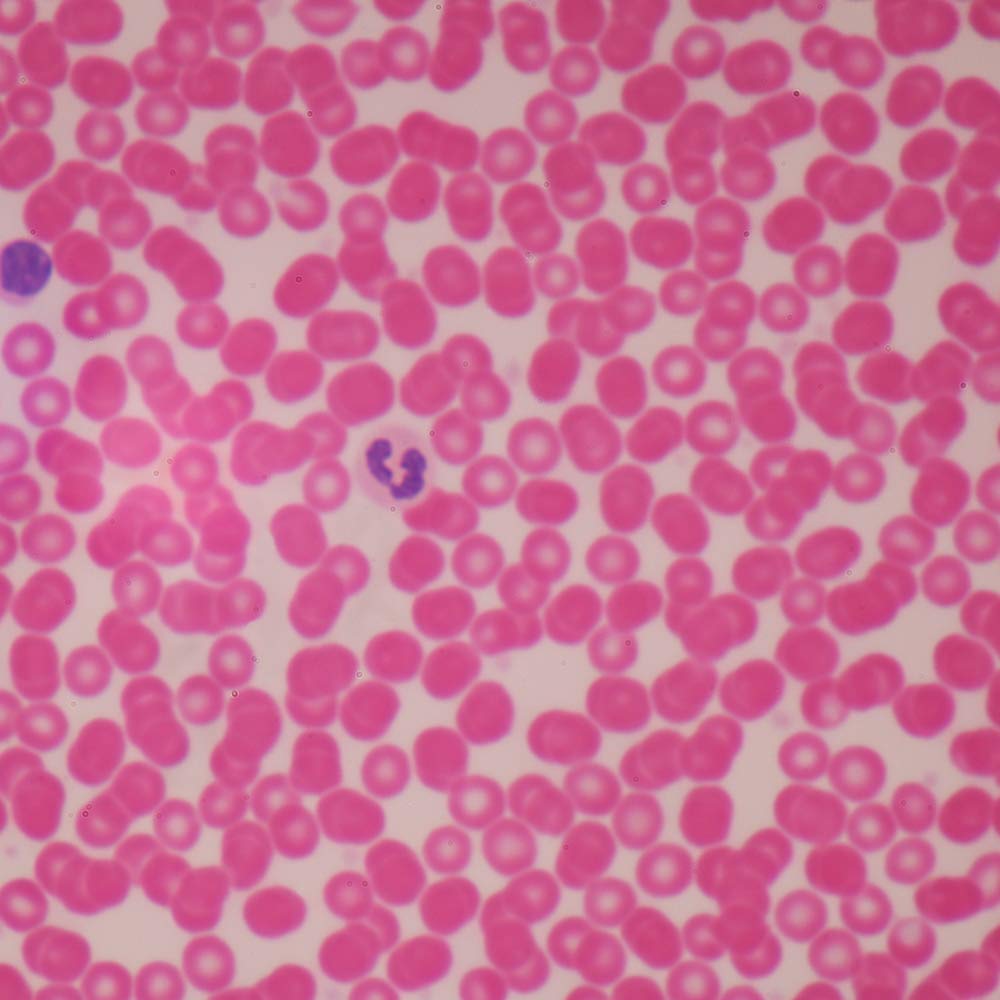
Light micrograph of a smear of human blood. The slide should be clean. Blood smear human wright s stain h6455 microscope at 1000x these images show erythrocytes red blood cells leukocytes white blood cells and platelets as they appear in a wright s stained blood smear. Blood smear human 5. C m ac m a a spreader slide is used to feather the drop of blood.
 Source: amazon.com
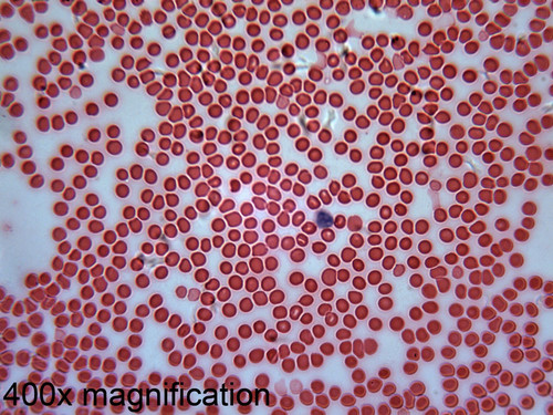
Blood smear erythrocytes red blood cells 100 x. Other of biology slide human blood smear slide chlamydomonas slide volvox slide and liver fluke slide offered by the national scientific suppliers chennai tamil nadu. Blood is an unusual connective tissue because it is normally in liquid form. Human blood smear showing redblood cells or erythrocytes and white blood. It consists of a fluid called plasma and cells formed elements that are suspended in the plasma.
 Source: amazon.com
Blood within 1 hr. C m ac m a a spreader slide is used to feather the drop of blood. C m ac m a blood smear preparation materials and supplies sample edta slides fixative buffer and stain diff quik water 4. Eosinophil basophil click on the any of the small images above to see a higher resolution image. Blood smear human 5.
 Source: slideshare.net
Cells will be heaped and piled close to the point were the drop of blood was placed on the slide. Where to look for cells in a blood smear the density of cells varies across the smear. Red blood cells leukemia platelets. 2 1 x 3 25 mm x 76 mm glass slides. Making and staining a blood smear a well made blood smear is a beauty to behold and likely to yield interesting and significant information for a research project.
 Source: sccollege.edu
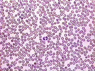
Blood smear human wright s stain h6455 microscope at 1000x these images show erythrocytes red blood cells leukocytes white blood cells and platelets as they appear in a wright s stained blood smear. Where to look for cells in a blood smear the density of cells varies across the smear. 3 packed in a plastic box human blood smear used to observe the cell composition of blood. Light micrograph of a smear of human blood. Red blood cells leukemia platelets.
 Source: austincc.edu
Of collection preparation of blood film. Place a small drop of blood or one side about 1 2 cm from one end. White blood cells appear shrunken and some types are difficult to distinguish from each other. It consists of a fluid called plasma and cells formed elements that are suspended in the plasma. C m ac m a blood smear preparation materials and supplies sample edta slides fixative buffer and stain diff quik water 4.
 Source: fishersci.com
Blood within 1 hr. A poor slide is a torment. The extra time and care taken during the field season will be rewarded later when the smears must be scanned and parasites identified and counted. White blood cells appear shrunken and some types are difficult to distinguish from each other. Preparation of blood smear.
If you find this site beneficial, please support us by sharing this posts to your favorite social media accounts like Facebook, Instagram and so on or you can also bookmark this blog page with the title human blood smear slide by using Ctrl + D for devices a laptop with a Windows operating system or Command + D for laptops with an Apple operating system. If you use a smartphone, you can also use the drawer menu of the browser you are using. Whether it’s a Windows, Mac, iOS or Android operating system, you will still be able to bookmark this website.
