Human blood smear wright
Home » Science Education » Human blood smear wrightHuman blood smear wright
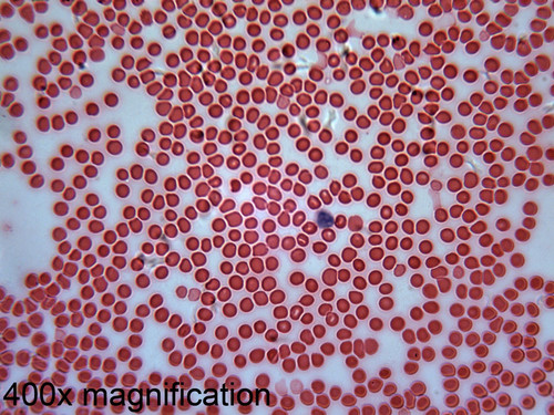
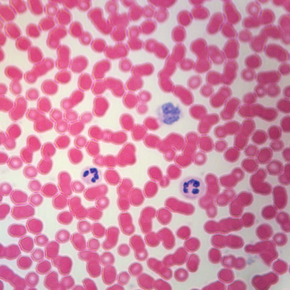
Human Blood Smear Wright. Blood is an unusual connective tissue because it is normally in liquid form. It is hoped that this paper will provide an aid to those who wish to employ the blood smear as a diagnostic tool. These images show erythrocytes red blood cells leukocytes white blood cells and platelets as they appear in a wright s stained blood smear. Making blood smears and in staining these smears with wright s stain.
 Blood From eugraph.com
Wright s stain is a hematologic stain that facilitates the differentiation of blood cell types. Blood within 1 hr. The three main blood cells that the test focuses on are. Human blood wright s stain smear. Wright s stain is a type of romanowsky stain which is commonly used in hematology laboratory for the routine staining of peripheral blood smears. Platelets are pinched off bits of cytoplasm of a bone marrow cell called a megakaryocyte enclosed in plasma membrane.
Wright s stain is a type of romanowsky stain which is commonly used in hematology laboratory for the routine staining of peripheral blood smears.
It consists of a fluid called plasma and cells formed elements that are suspended in the plasma. It is used primarily to stain peripheral blood smears urine samples and bone marrow aspirates which are examined under a light microscope. Based on the shape and size of the nucleus in the white blood cells students can classify them as neutrophils eosinophils basophils lymphocytes or monocytes. Wright s stain is a type of romanowsky stain which is commonly used in hematology laboratory for the routine staining of peripheral blood smears. Blood is an unusual connective tissue because it is normally in liquid form. Making blood smears and in staining these smears with wright s stain.
 Source: amazon.com
Blood is an unusual connective tissue because it is normally in liquid form. Blood is an unusual connective tissue because it is normally in liquid form. Blood within 1 hr. Wright s stain is a hematologic stain that facilitates the differentiation of blood cell types. The slide from which this image was prepared was a blood smear it was made by putting a drop of blood on one end of a slide and using a second slide to spread the blood.
 Source: geiselmed.dartmouth.edu
Platelets contain various substances that contribute to blood clotting. Of collection preparation of blood film. A blood smear is a blood test used to look for abnormalities in blood cells. Preparation of blood smear. It is classically a mixture of eosin red and methylene blue dyes.
 Source: eugraph.com
The slide should be clean. In cytogenetics it is used to stain. Platelets are pinched off bits of cytoplasm of a bone marrow cell called a megakaryocyte enclosed in plasma membrane. Platelets contain various substances that contribute to blood clotting. This blood slide is stained with wright s stain to make the erythrocytes leukocytes platelets well differentiated.
 Source: researchgate.net
The three main blood cells that the test focuses on are. The mechanism of action of wright s stain is also discussed. Of collection preparation of blood film. It consists of a fluid called plasma and cells formed elements that are suspended in the plasma. Making blood smears and in staining these smears with wright s stain.
 Source: geiselmed.dartmouth.edu
The three main blood cells that the test focuses on are. The three main blood cells that the test focuses on are. The slide from which this image was prepared was a blood smear it was made by putting a drop of blood on one end of a slide and using a second slide to spread the blood. For a slide which highlights chromosomes our giemsa stain slide is ideal. It consists of a fluid called plasma and cells formed elements that are suspended in the plasma.
 Source: amazon.com
In cytogenetics it is used to stain. The slide should be clean. Blood within 1 hr. Human blood wright s stain smear. It is used primarily to stain peripheral blood smears urine samples and bone marrow aspirates which are examined under a light microscope.
 Source: fishersci.com
Save to your sharing list ask a question about this product 0 description. Without delay place a spreader at an angle of 45 from the slide and move it back to make contact with. For a slide which highlights chromosomes our giemsa stain slide is ideal. Wright s stain is a hematologic stain that facilitates the differentiation of blood cell types. It is hoped that this paper will provide an aid to those who wish to employ the blood smear as a diagnostic tool.
 Source: eugraph.com
It is also used for staining bone marrow aspirates urine samples and to demonstrate malarial parasites in blood smears. The slide should be clean. Save to your sharing list ask a question about this product 0 description. Of collection preparation of blood film. In cytogenetics it is used to stain.
 Source: geiselmed.dartmouth.edu
Blood within 1 hr. A blood smear is a blood test used to look for abnormalities in blood cells. For a slide which highlights chromosomes our giemsa stain slide is ideal. The slide should be clean. Save to your sharing list ask a question about this product 0 description.
 Source: quizlet.com
It is used primarily to stain peripheral blood smears urine samples and bone marrow aspirates which are examined under a light microscope. Blood within 1 hr. In cytogenetics it is used to stain. Wright s stain is a type of romanowsky stain which is commonly used in hematology laboratory for the routine staining of peripheral blood smears. It is also used for staining bone marrow aspirates urine samples and to demonstrate malarial parasites in blood smears.
 Source: homesciencetools.com
Save to your sharing list ask a question about this product 0 description. Save to your sharing list ask a question about this product 0 description. A blood smear is a blood test used to look for abnormalities in blood cells. Platelets are pinched off bits of cytoplasm of a bone marrow cell called a megakaryocyte enclosed in plasma membrane. Leukocytes white blood cells are larger cells with a purple nucleus.
 Source: fishersci.com
It is used primarily to stain peripheral blood smears urine samples and bone marrow aspirates which are examined under a light microscope. Place a small drop of blood or one side about 1 2 cm from one end. Human blood wright s stain smear. Blood is an unusual connective tissue because it is normally in liquid form. Platelets are pinched off bits of cytoplasm of a bone marrow cell called a megakaryocyte enclosed in plasma membrane.
 Source: quizlet.com
Wright s stain is a type of romanowsky stain which is commonly used in hematology laboratory for the routine staining of peripheral blood smears. This blood slide is stained with wright s stain to make the erythrocytes leukocytes platelets well differentiated. It is classically a mixture of eosin red and methylene blue dyes. Of collection preparation of blood film. Red cells which carry oxygen throughout your body.
 Source: amazon.com
A blood smear is a blood test used to look for abnormalities in blood cells. This blood slide is stained with wright s stain to make the erythrocytes leukocytes platelets well differentiated. In cytogenetics it is used to stain. The blood smear is stained with wright s stain allowing students to easily distinguish between red and white blood cells. Platelets are pinched off bits of cytoplasm of a bone marrow cell called a megakaryocyte enclosed in plasma membrane.
 Source: eugraph.com
Wright s stain is named for james homer wright who devised the stain in 1902 based on a modification of romanowsky stain. It is classically a mixture of eosin red and methylene blue dyes. Making the blood smear before any stained smear can be used for a diagnosis. Save to your sharing list ask a question about this product 0 description. Wright s stain is a hematologic stain that facilitates the differentiation of blood cell types.
If you find this site good, please support us by sharing this posts to your favorite social media accounts like Facebook, Instagram and so on or you can also save this blog page with the title human blood smear wright by using Ctrl + D for devices a laptop with a Windows operating system or Command + D for laptops with an Apple operating system. If you use a smartphone, you can also use the drawer menu of the browser you are using. Whether it’s a Windows, Mac, iOS or Android operating system, you will still be able to bookmark this website.
