Human eye labelled
Home » Science Education » Human eye labelledHuman eye labelled
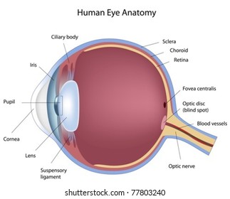
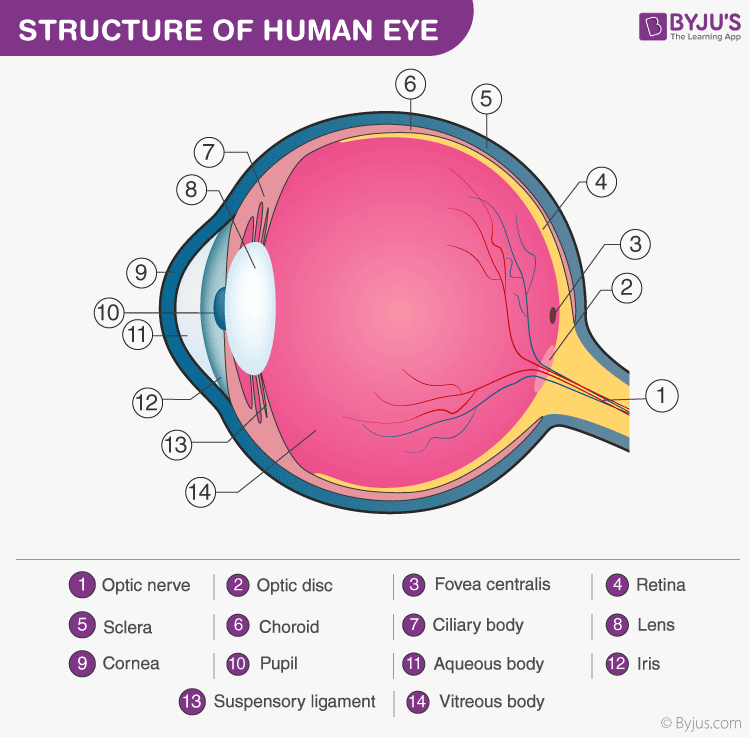
Human Eye Labelled. Light is focused primarily by the cornea the clear front surface of the eye which acts like a camera lens. A human eye is roughly 2 3 cm in diameter and is almost a spherical ball filled with some fluid. The front transparent part of the sclera is called cornea. Hence it does not possess a perfect spherical shape.
 Human Eye Diagram Images Stock Photos Vectors Shutterstock From shutterstock.com
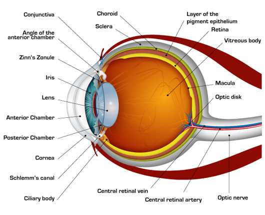
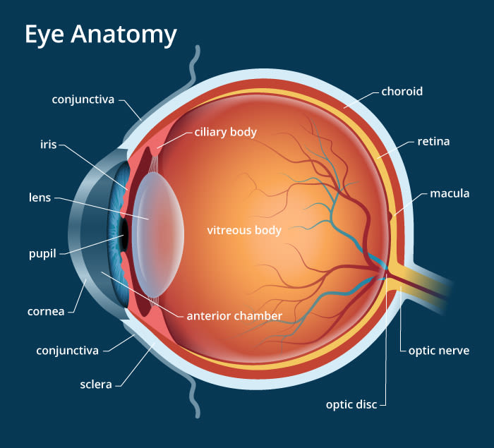
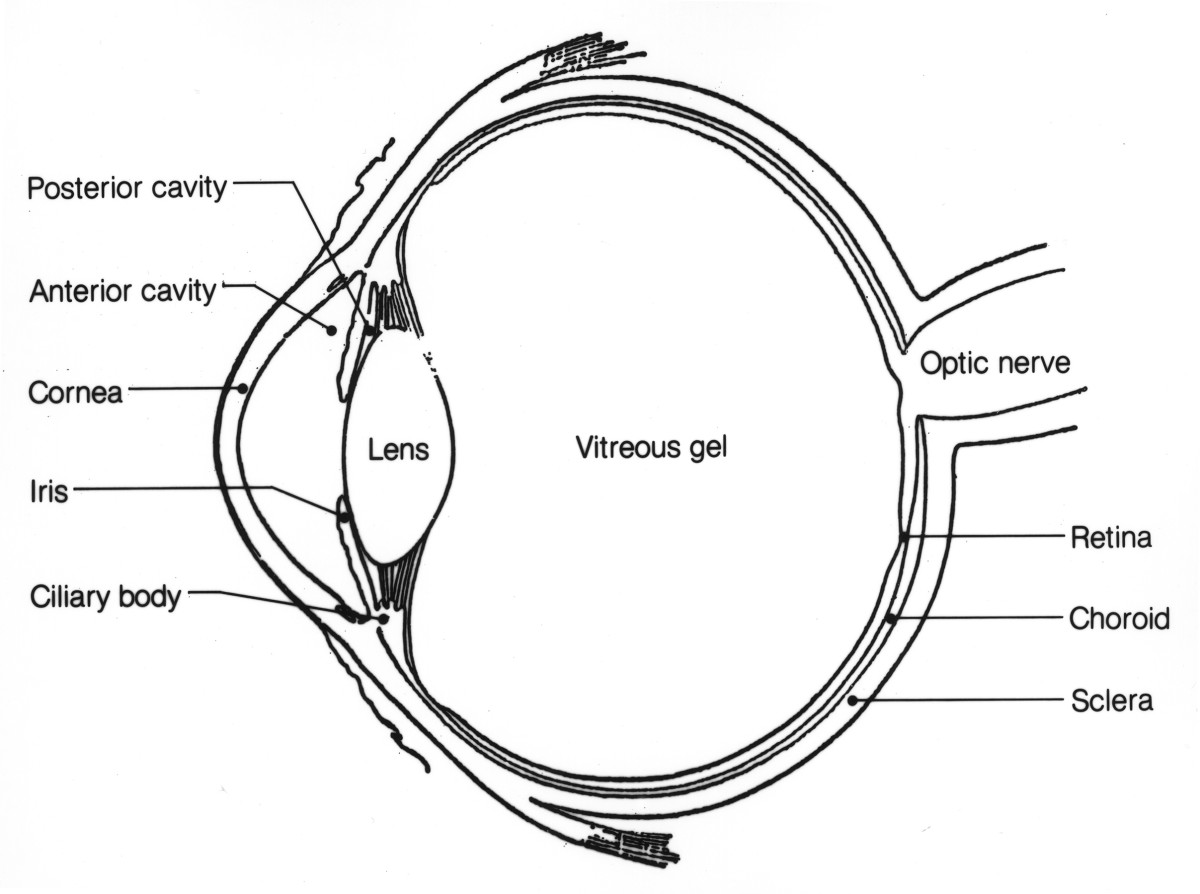
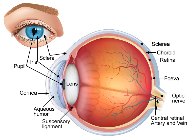
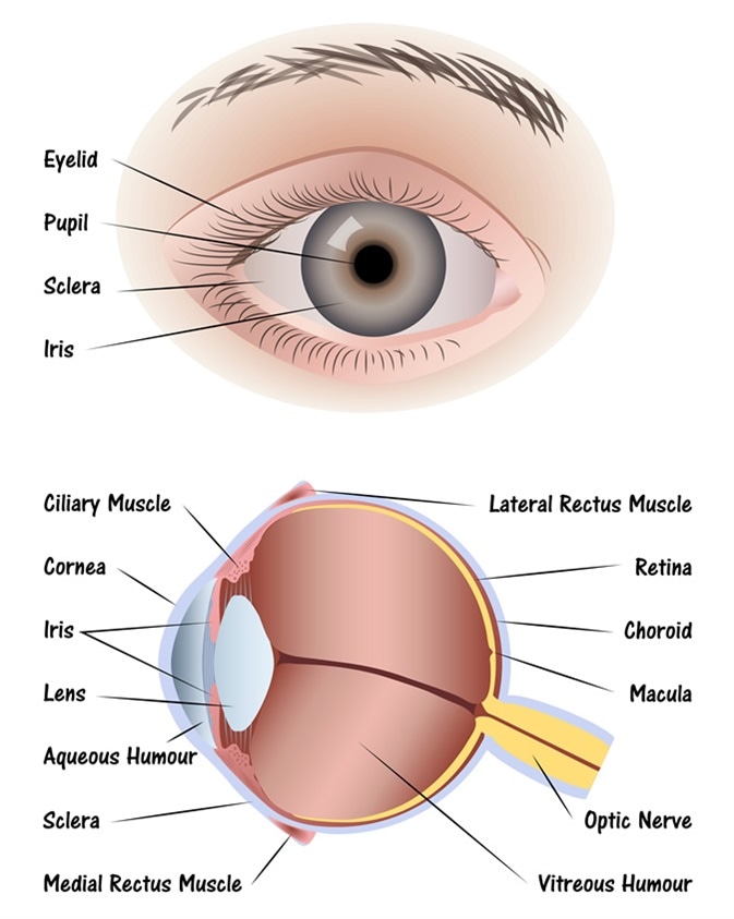
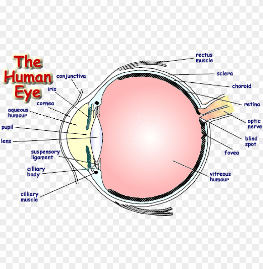
Multiple genes inherited from each. The front transparent part of the sclera is called cornea. Hence it does not possess a perfect spherical shape. The diagrams below show cross sections of the human eyeball. Human eye specialized sense organ in humans that is capable of receiving visual images which are relayed to the brain. The different parts of the eye allow the body to take in light and perceive objects around us in the proper.
It is the outer covering a protective tough white layer called the sclera white part of the eye.
It consists of the following parts. Behind the anterior chamber is the eye s iris the colored part of the eye and the dark hole in the middle called the pupil. It is enclosed within the eye sockets in the skull and is anchored down by muscles within the sockets. As we journey through the different structures refer to the diagrams to quickly digest the content on this page. Eye color is created by the amount and type of pigment in your iris. Rod and cone cells in the retina are photoreceptive cells which are able to detect visible light and convey this information to the brain eyes signal information which is used by the brain to elicit the perception of color shape depth movement and other features.
 Source: pinterest.com
It is enclosed within the eye sockets in the skull and is anchored down by muscles within the sockets. The diagrams below show cross sections of the human eyeball. Hence it does not possess a perfect spherical shape. Parts of human eye and their functions understanding the different parts of our eye can help you understand how you see and what you can do to help keep the eye functioning properly. Multiple genes inherited from each.
 Source: familyconnect.org
In a number of ways the human eye works much like a digital camera. Eye color is created by the amount and type of pigment in your iris. Let s take a closer look at each of these. This article explores the anatomy of the eye looking at the different structures of the human eye and their function. Structure of human eye.
 Source: byjus.com
What you want to interpret as a major part of the human eye is somewhat up to the individual but in general there are seven parts of the human eye. Parts of human eye and their functions understanding the different parts of our eye can help you understand how you see and what you can do to help keep the eye functioning properly. The anatomy of the eye includes auxiliary structures such as the bony eye socket and extraocular muscles as well as the structures of the eye itself such as the lens and the retina. The cornea the pupil the iris the lens the vitreous humor the retina and the sclera. The eye is always producing aqueous humor.
 Source: researchgate.net
The macula is a small extra sensitive area in the retina that gives you central vision. The human eye is composed of many different parts that work together to interpret the world around us. The diagrams below show cross sections of the human eyeball. The eye is always producing aqueous humor. What you want to interpret as a major part of the human eye is somewhat up to the individual but in general there are seven parts of the human eye.
 Source: pinterest.com
The human eye is a paired sense organ that reacts to light and allows vision. The human eye is composed of many different parts that work together to interpret the world around us. The human eye is a paired sense organ that reacts to light and allows vision. The human eye is a roughly spherical organ responsible for perceiving visual stimuli. Human eye specialized sense organ in humans that is capable of receiving visual images which are relayed to the brain.
 Source: nkcf.org
Parts of human eye and their functions understanding the different parts of our eye can help you understand how you see and what you can do to help keep the eye functioning properly. Behind the anterior chamber is the eye s iris the colored part of the eye and the dark hole in the middle called the pupil. Light enters the eye through the cornea. The anatomy of the eye includes auxiliary structures such as the bony eye socket and extraocular muscles as well as the structures of the eye itself such as the lens and the retina. The macula is a small extra sensitive area in the retina that gives you central vision.
 Source: shutterstock.com
Anatomically the eye comprises two components fused into one. Rod and cone cells in the retina are photoreceptive cells which are able to detect visible light and convey this information to the brain eyes signal information which is used by the brain to elicit the perception of color shape depth movement and other features. The macula is a small extra sensitive area in the retina that gives you central vision. Light is focused primarily by the cornea the clear front surface of the eye which acts like a camera lens. It consists of the following parts.
 Source: allaboutvision.com
A human eye is roughly 2 3 cm in diameter and is almost a spherical ball filled with some fluid. Light is focused primarily by the cornea the clear front surface of the eye which acts like a camera lens. Rod and cone cells in the retina are photoreceptive cells which are able to detect visible light and convey this information to the brain eyes signal information which is used by the brain to elicit the perception of color shape depth movement and other features. The macula is a small extra sensitive area in the retina that gives you central vision. Parts of human eye and their functions understanding the different parts of our eye can help you understand how you see and what you can do to help keep the eye functioning properly.
 Source: owlcation.com
It is the outer covering a protective tough white layer called the sclera white part of the eye. It is enclosed within the eye sockets in the skull and is anchored down by muscles within the sockets. In a number of ways the human eye works much like a digital camera. It is the outer covering a protective tough white layer called the sclera white part of the eye. The different parts of the eye allow the body to take in light and perceive objects around us in the proper.
Source: sarthaks.com
The human eye is a roughly spherical organ responsible for perceiving visual stimuli. The cornea the pupil the iris the lens the vitreous humor the retina and the sclera. The eye is part of the sensory nervous system. Muscles in the iris dilate widen or constrict narrow the. The eye is one of the most complex parts of the body.
 Source: topperlearning.com
Light enters the eye through the cornea. The anatomy of the eye includes auxiliary structures such as the bony eye socket and extraocular muscles as well as the structures of the eye itself such as the lens and the retina. Muscles in the iris dilate widen or constrict narrow the. Structure of human eye. The different parts of the eye allow the body to take in light and perceive objects around us in the proper.
 Source: news-medical.net
The macula is a small extra sensitive area in the retina that gives you central vision. The eye is one of the most complex parts of the body. Structure of human eye. A human eye is roughly 2 3 cm in diameter and is almost a spherical ball filled with some fluid. In a number of ways the human eye works much like a digital camera.
 Source: thoughtco.com
Rod and cone cells in the retina are photoreceptive cells which are able to detect visible light and convey this information to the brain eyes signal information which is used by the brain to elicit the perception of color shape depth movement and other features. The macula is a small extra sensitive area in the retina that gives you central vision. This article explores the anatomy of the eye looking at the different structures of the human eye and their function. Muscles in the iris dilate widen or constrict narrow the. It is enclosed within the eye sockets in the skull and is anchored down by muscles within the sockets.
 Source: boozmanhof.com
The eye is one of the most complex parts of the body. The iris of the eye functions like the diaphragm of a camera controlling the amount of light reaching the back of the eye by automatically adjusting the size of the. Light enters the eye through the cornea. What you want to interpret as a major part of the human eye is somewhat up to the individual but in general there are seven parts of the human eye. This article explores the anatomy of the eye looking at the different structures of the human eye and their function.
 Source: toppng.com
Multiple genes inherited from each. The human eye is a roughly spherical organ responsible for perceiving visual stimuli. Eye color is created by the amount and type of pigment in your iris. The iris of the eye functions like the diaphragm of a camera controlling the amount of light reaching the back of the eye by automatically adjusting the size of the. The diagrams below show cross sections of the human eyeball.
If you find this site serviceableness, please support us by sharing this posts to your own social media accounts like Facebook, Instagram and so on or you can also bookmark this blog page with the title human eye labelled by using Ctrl + D for devices a laptop with a Windows operating system or Command + D for laptops with an Apple operating system. If you use a smartphone, you can also use the drawer menu of the browser you are using. Whether it’s a Windows, Mac, iOS or Android operating system, you will still be able to bookmark this website.
