Inferior vena cava diagram
Home » Science Education » Inferior vena cava diagramInferior vena cava diagram
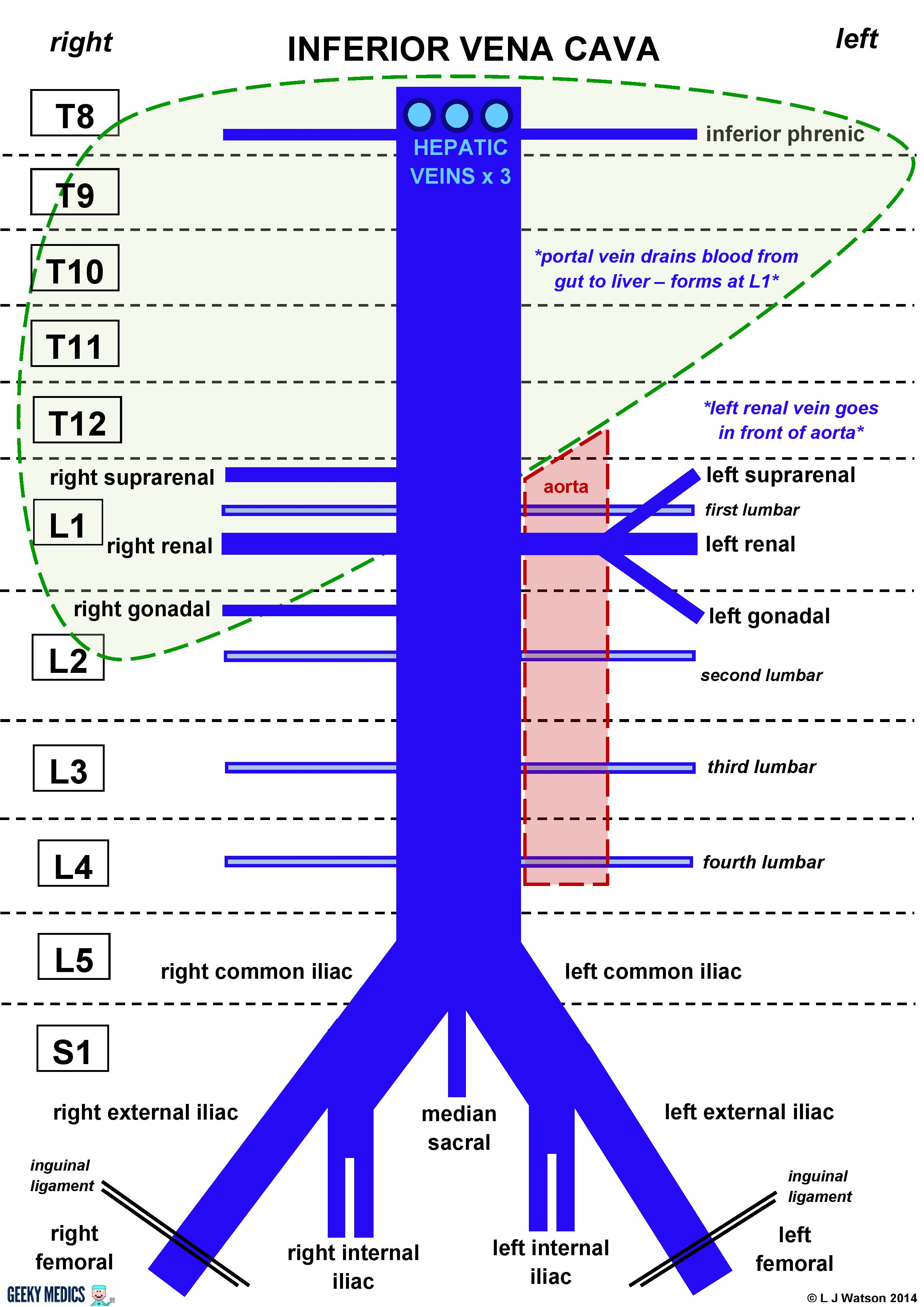
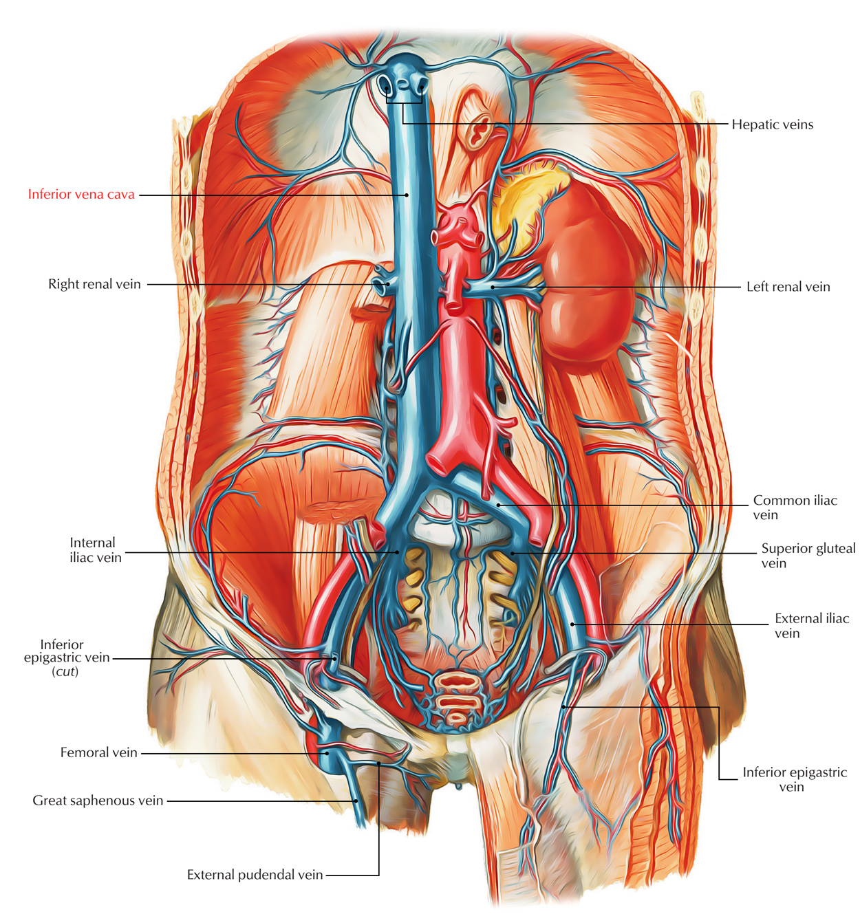
Inferior Vena Cava Diagram. There are several key points to take away from this diagram. Although the vena cava is very large in diameter its walls are incredibly thin due to the low pressure exerted by venous blood. In this image you will find hepatic veins inferior phrenic vein portal vein left renal vein left suprarenal vein left gonadal vein right gonadal vein right renal vein in it. You may also find right suprarenal vein aorta left common iliac vein right common iliac vein left external iliac vein median sacral.
 Tributaries Of Inferior Vena Cava Diagram Quizlet From quizlet.com
Inferior vena cava ivc key points. It is formed by the joining of the right and the left common iliac veins usually at the level of the fifth lumbar vertebra. The inferior vena cava is the lower of the two venae cavae the two large veins that carry deoxygenated blood from the body to the right atrium of the heart. 3 anterior visceral tributaries three hepatic 3 lateral visceral tributaries suprarenal renal gonadal 5 lateral abdominal wall tributaries inferior phrenic and four lumbar 3 veins of origin two common iliac and the median sacral. The inferior vena cava carries blood from the lower half of the body whilst. The inferior vena cava is the largest vein in the human body.
The inferior vena cava is the largest vein in the human body.
The inferior vena cava carries blood from the lower half of the body whilst. Although the vena cava is very large in diameter its walls are incredibly thin due to the low pressure exerted by venous blood. 3 anterior visceral tributaries three hepatic 3 lateral visceral tributaries suprarenal renal gonadal 5 lateral abdominal wall tributaries inferior phrenic and four lumbar 3 veins of origin two common iliac and the median sacral. Inferior vena cava ivc key points. It collects blood from veins serving the tissues inferior to the heart and returns this blood to the right atrium of the heart. The inferior vena cava is a large vein that carries de oxygenated blood from the lower body to the heart.
 Source: pinterest.com.mx
It is formed by the joining of the right and the left common iliac veins usually at the level of the fifth lumbar vertebra. The ivc a single right sided vessel in 97 of individuals returns blood from all structures below the diaphragm to the right atrium of the heart. It is formed by the confluence of the two common iliac veins at the level of the fifth lumbar vertebra just to the right of midline. The inferior vena cava is the largest vein in the human body. It collects blood from veins serving the tissues inferior to the heart and returns this blood to the right atrium of the heart.
 Source: researchgate.net
Inferior vena cava ivc key points. Although the vena cava is very large in diameter its walls are incredibly thin due to the low pressure exerted by venous blood. It is formed by the confluence of the two common iliac veins at the level of the fifth lumbar vertebra just to the right of midline. The ivc a single right sided vessel in 97 of individuals returns blood from all structures below the diaphragm to the right atrium of the heart. You may also find right suprarenal vein aorta left common iliac vein right common iliac vein left external iliac vein median sacral.
 Source: researchgate.net
3 anterior visceral tributaries three hepatic 3 lateral visceral tributaries suprarenal renal gonadal 5 lateral abdominal wall tributaries inferior phrenic and four lumbar 3 veins of origin two common iliac and the median sacral. Inferior vena cava diagram. The inferior vena cava is also referred to as the posterior vena cava. You may also find right suprarenal vein aorta left common iliac vein right common iliac vein left external iliac vein median sacral. In this image you will find hepatic veins inferior phrenic vein portal vein left renal vein left suprarenal vein left gonadal vein right gonadal vein right renal vein in it.
 Source: en.wikipedia.org
There are several key points to take away from this diagram. It collects blood from veins serving the tissues inferior to the heart and returns this blood to the right atrium of the heart. It is formed by the confluence of the two common iliac veins at the level of the fifth lumbar vertebra just to the right of midline. Inferior vena cava diagram. In this image you will find hepatic veins inferior phrenic vein portal vein left renal vein left suprarenal vein left gonadal vein right gonadal vein right renal vein in it.
 Source: quizlet.com
Although the vena cava is very large in diameter its walls are incredibly thin due to the low pressure exerted by venous blood. The inferior vena cava is the lower of the two venae cavae the two large veins that carry deoxygenated blood from the body to the right atrium of the heart. Inferior vena cava ivc key points. Although the vena cava is very large in diameter its walls are incredibly thin due to the low pressure exerted by venous blood. The inferior vena cava is a large vein that carries the deoxygenated blood from the lower and middle body into the right atrium of the heart.
 Source: britannica.com
3 anterior visceral tributaries three hepatic 3 lateral visceral tributaries suprarenal renal gonadal 5 lateral abdominal wall tributaries inferior phrenic and four lumbar 3 veins of origin two common iliac and the median sacral. You may also find right suprarenal vein aorta left common iliac vein right common iliac vein left external iliac vein median sacral. 3 anterior visceral tributaries three hepatic 3 lateral visceral tributaries suprarenal renal gonadal 5 lateral abdominal wall tributaries inferior phrenic and four lumbar 3 veins of origin two common iliac and the median sacral. The inferior vena cava is a large vein that carries de oxygenated blood from the lower body to the heart. Inferior vena cava ivc key points.
 Source: researchgate.net
There are several key points to take away from this diagram. The inferior vena cava carries blood from the lower half of the body whilst. The inferior vena cava is a large vein that carries the deoxygenated blood from the lower and middle body into the right atrium of the heart. It is formed by the confluence of the two common iliac veins at the level of the fifth lumbar vertebra just to the right of midline. The inferior vena cava is the largest vein in the human body.
 Source: radiologykey.com
The ivc a single right sided vessel in 97 of individuals returns blood from all structures below the diaphragm to the right atrium of the heart. It is formed by the confluence of the two common iliac veins at the level of the fifth lumbar vertebra just to the right of midline. The inferior vena cava is also referred to as the posterior vena cava. The inferior vena cava is the largest vein in the human body. There are several key points to take away from this diagram.
 Source: kenhub.com
There are several key points to take away from this diagram. Although the vena cava is very large in diameter its walls are incredibly thin due to the low pressure exerted by venous blood. 3 anterior visceral tributaries three hepatic 3 lateral visceral tributaries suprarenal renal gonadal 5 lateral abdominal wall tributaries inferior phrenic and four lumbar 3 veins of origin two common iliac and the median sacral. There are several key points to take away from this diagram. It is formed by the joining of the right and the left common iliac veins usually at the level of the fifth lumbar vertebra.
 Source: quizlet.com
The inferior vena cava carries blood from the lower half of the body whilst. The inferior vena cava is a large vein that carries de oxygenated blood from the lower body to the heart. There are several key points to take away from this diagram. The inferior vena cava is the largest vein in the human body. The ivc a single right sided vessel in 97 of individuals returns blood from all structures below the diaphragm to the right atrium of the heart.
 Source: researchgate.net
You may also find right suprarenal vein aorta left common iliac vein right common iliac vein left external iliac vein median sacral. 3 anterior visceral tributaries three hepatic 3 lateral visceral tributaries suprarenal renal gonadal 5 lateral abdominal wall tributaries inferior phrenic and four lumbar 3 veins of origin two common iliac and the median sacral. The inferior vena cava is the largest vein in the human body. The inferior vena cava is the lower of the two venae cavae the two large veins that carry deoxygenated blood from the body to the right atrium of the heart. The inferior vena cava carries blood from the lower half of the body whilst.
 Source: geekymedics.com
Inferior vena cava ivc key points. Inferior vena cava ivc key points. The inferior vena cava is a large vein that carries the deoxygenated blood from the lower and middle body into the right atrium of the heart. You may also find right suprarenal vein aorta left common iliac vein right common iliac vein left external iliac vein median sacral. Although the vena cava is very large in diameter its walls are incredibly thin due to the low pressure exerted by venous blood.
 Source: sciencedirect.com
It collects blood from veins serving the tissues inferior to the heart and returns this blood to the right atrium of the heart. You may also find right suprarenal vein aorta left common iliac vein right common iliac vein left external iliac vein median sacral. The inferior vena cava is the largest vein in the human body. It is formed by the confluence of the two common iliac veins at the level of the fifth lumbar vertebra just to the right of midline. The inferior vena cava carries blood from the lower half of the body whilst.
 Source: earthslab.com
3 anterior visceral tributaries three hepatic 3 lateral visceral tributaries suprarenal renal gonadal 5 lateral abdominal wall tributaries inferior phrenic and four lumbar 3 veins of origin two common iliac and the median sacral. The inferior vena cava is also referred to as the posterior vena cava. The inferior vena cava is a large vein that carries the deoxygenated blood from the lower and middle body into the right atrium of the heart. Although the vena cava is very large in diameter its walls are incredibly thin due to the low pressure exerted by venous blood. The inferior vena cava carries blood from the lower half of the body whilst.
 Source: en.wikipedia.org
In this image you will find hepatic veins inferior phrenic vein portal vein left renal vein left suprarenal vein left gonadal vein right gonadal vein right renal vein in it. Inferior vena cava ivc key points. In this image you will find hepatic veins inferior phrenic vein portal vein left renal vein left suprarenal vein left gonadal vein right gonadal vein right renal vein in it. There are several key points to take away from this diagram. The inferior vena cava is the lower of the two venae cavae the two large veins that carry deoxygenated blood from the body to the right atrium of the heart.
If you find this site helpful, please support us by sharing this posts to your own social media accounts like Facebook, Instagram and so on or you can also bookmark this blog page with the title inferior vena cava diagram by using Ctrl + D for devices a laptop with a Windows operating system or Command + D for laptops with an Apple operating system. If you use a smartphone, you can also use the drawer menu of the browser you are using. Whether it’s a Windows, Mac, iOS or Android operating system, you will still be able to bookmark this website.
