Neuron microscope slide
Home » Science Education » Neuron microscope slideNeuron microscope slide
Neuron Microscope Slide. What is a neuron br univeristy of saint thomas br 2. Always handle the slides with care as they can easily be shattered or scratched. Follow the following steps in order to observe different parts of the neuron. Br the process by which a neuron carries messages from one part of the brain to another is through what is called an electrochemical process.
 Nervous System Lab From medcell.med.yale.edu
Perform a first hand investigation using stained prepared slides and or electron micrographs to gather informat. Follow the following steps in order to observe different parts of the neuron. For more anatomy content please follow us and visit our website. Always handle the slides with care as they can easily be shattered or scratched. A brief discussion of a microscope slide containing a neuron smear with emphasis on anatomy and some function. Br the electro part happens in the neuron itself.
Under the microscope procedure.
What is a neuron 1. What is a neuron 1. Locate a large neuron and move it to the center of your field of view. Never drop slides or slide covers and set them down only on clean countertops. Br the chemical part happens at a junction. Supporting cells do not conduct nerve impulses but they perform many other functions for the nerve tissue.
 Source: medsci.indiana.edu
Microscope slides and slide covers are small and delicate. Covers the hsc biology syllabus dot point. Microscope slides and slide covers are small and delicate. Br the process by which a neuron carries messages from one part of the brain to another is through what is called an electrochemical process. Always handle the slides with care as they can easily be shattered or scratched.
 Source: medcell.med.yale.edu
What is a neuron 1. Securing the slide on the stage using the stage clips using the fine adjustment knob to. Locate a large neuron and move it to the center of your field of view. Supporting cells do not conduct nerve impulses but they perform many other functions for the nerve tissue. Microscope slides and slide covers are small and delicate.
 Source: blades-bio.co.uk
Follow the following steps in order to observe different parts of the neuron. Never drop slides or slide covers and set them down only on clean countertops. A neuron is a cell in the body that is specialized to carry messages. Always handle the slides with care as they can easily be shattered or scratched. A brief discussion of a microscope slide containing a neuron smear with emphasis on anatomy and some function.
 Source: medcell.med.yale.edu
Like other cells neurons can be observed under a light microscope. We are pleased to provide you with the picture named neuron anatomy in detail and microscope view we hope this picture neuron anatomy in detail and microscope view can help you study and research. Which of the following is the first step in using a light microscope to observe a prepared slide of neurons. Supporting cells do not conduct nerve impulses but they perform many other functions for the nerve tissue. A brief discussion of a microscope slide containing a neuron smear with emphasis on anatomy and some function.
 Source: austincc.edu
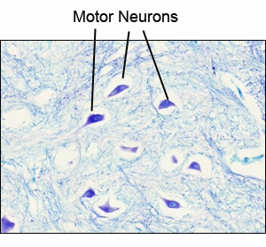
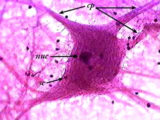
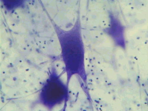
The images on this page were made from a slide called a motor neuron smear. Like other cells neurons can be observed under a light microscope. What is a neuron br univeristy of saint thomas br 2. Niemann pick disease accumulation of sphingomyelin in lysosomes. Always handle the slides with care as they can easily be shattered or scratched.
 Source: dartmouth.edu
Br the chemical part happens at a junction. What is a neuron br univeristy of saint thomas br 2. Ganglion cells in tay sachs disease a under the light microscope a large neuron has obvious lipid vacuolation b a portion of a neuron under the electron microscope shows prominent lysosomes withwhorled configurations 79. Covers the hsc biology syllabus dot point. Under the microscope procedure.
 Source: medcell.med.yale.edu
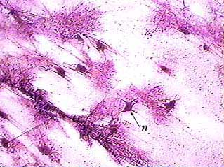
Br the electro part happens in the neuron itself. Br the chemical part happens at a junction. What is a neuron 1. Neurons n are the ones that generate and conduct nerve impulses. Under the microscope procedure.
 Source: austincc.edu
Supporting cells do not conduct nerve impulses but they perform many other functions for the nerve tissue. Niemann pick disease accumulation of sphingomyelin in lysosomes. For more anatomy content please follow us and visit our website. Neurons n are the ones that generate and conduct nerve impulses. Covers the hsc biology syllabus dot point.
 Source: faculty.une.edu
Perform a first hand investigation using stained prepared slides and or electron micrographs to gather informat. Iodine and methylene blue are poisonous and should never be ingested. Locate a large neuron and move it to the center of your field of view. What is a neuron 1. Covers the hsc biology syllabus dot point.
 Source: eugraph.com
A brief discussion of a microscope slide containing a neuron smear with emphasis on anatomy and some function. Perform a first hand investigation using stained prepared slides and or electron micrographs to gather informat. Which of the following is the first step in using a light microscope to observe a prepared slide of neurons. The images on this page were made from a slide called a motor neuron smear. Ganglion cells in tay sachs disease a under the light microscope a large neuron has obvious lipid vacuolation b a portion of a neuron under the electron microscope shows prominent lysosomes withwhorled configurations 79.
 Source: medcell.med.yale.edu
Br the chemical part happens at a junction. Never drop slides or slide covers and set them down only on clean countertops. A brief discussion of a microscope slide containing a neuron smear with emphasis on anatomy and some function. What is a neuron 1. Always handle the slides with care as they can easily be shattered or scratched.
 Source: austincc.edu
Follow the following steps in order to observe different parts of the neuron. Locate a large neuron and move it to the center of your field of view. Supporting cells do not conduct nerve impulses but they perform many other functions for the nerve tissue. Neurons n are the ones that generate and conduct nerve impulses. Securing the slide on the stage using the stage clips using the fine adjustment knob to.
 Source: pinterest.com
Follow the following steps in order to observe different parts of the neuron. Covers the hsc biology syllabus dot point. Always handle the slides with care as they can easily be shattered or scratched. Follow the following steps in order to observe different parts of the neuron. Switch to the low power lens 10x get the neuron into focus and move it to the center of your field of view.
 Source: kenhub.com
What is a neuron br univeristy of saint thomas br 2. Covers the hsc biology syllabus dot point. Locate a large neuron and move it to the center of your field of view. A neuron is a cell in the body that is specialized to carry messages. Follow the following steps in order to observe different parts of the neuron.
 Source: homesciencetools.com
We are pleased to provide you with the picture named neuron anatomy in detail and microscope view we hope this picture neuron anatomy in detail and microscope view can help you study and research. For more anatomy content please follow us and visit our website. Which of the following is the first step in using a light microscope to observe a prepared slide of neurons. Like other cells neurons can be observed under a light microscope. Br the process by which a neuron carries messages from one part of the brain to another is through what is called an electrochemical process.
If you find this site good, please support us by sharing this posts to your favorite social media accounts like Facebook, Instagram and so on or you can also save this blog page with the title neuron microscope slide by using Ctrl + D for devices a laptop with a Windows operating system or Command + D for laptops with an Apple operating system. If you use a smartphone, you can also use the drawer menu of the browser you are using. Whether it’s a Windows, Mac, iOS or Android operating system, you will still be able to bookmark this website.
