Sheep brain dissection diagram
Home » Science Education » Sheep brain dissection diagramSheep brain dissection diagram
Sheep Brain Dissection Diagram. See diagram below and how this may influence the color differences you see. Place the brain with the curved top side of the. Learn vocabulary terms and more with flashcards games and other study tools. The sheep brain is quite similar to the human brain except for proportion.
 Sheep Brain Dissection Biology4friends From biology4friends.org
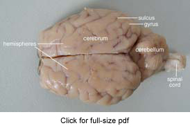
Start studying sheep brain dissection labeled. Sheep brain dissection. The sheep has a smaller cerebrum. Place the brain with the curved top side of the cerebrum facing up. Place the brain with the curved top side of the. Separate the two halves hemispheres.
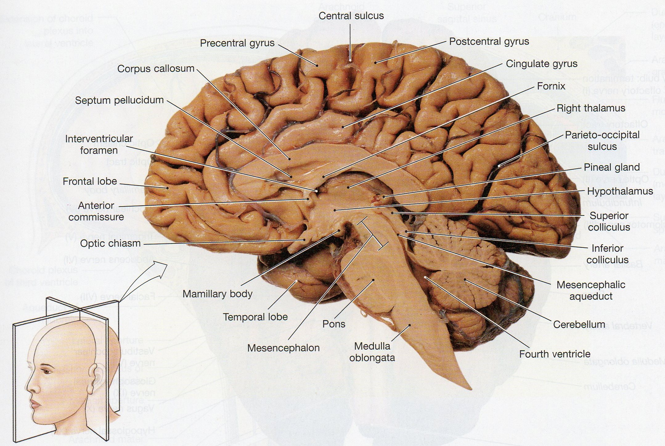
Also the sheep brain is oriented anterior to posterior more horizontally while the human brain is oriented superior to interior more vertically materials.
Place the brain with the curved top side of the cerebrum facing up. Separate the two halves hemispheres. The sheep has a smaller cerebrum. Place the brain with the curved top side of the. Cerebellum spinal cord medulla and. Separating the two.
 Source: biology4friends.org
Place the brain with the curved top side of the. Learn vocabulary terms and more with flashcards games and other study tools. Sheep brain dissection. And going down through the. Separate the two halves of the brain and lay them with the inside facing up.
 Source: courses.lumenlearning.com
Sheep brain dissection michigan state university neuroscience program brain bee enrichment workshop october 6 2012. Dissection tools and tray lab gloves preserved sheep brain. Slice through the brain along the center line starting at the. See diagram below and how this may influence the color differences you see. Start studying sheep brain dissection labeled.
 Source: learning-center.homesciencetools.com
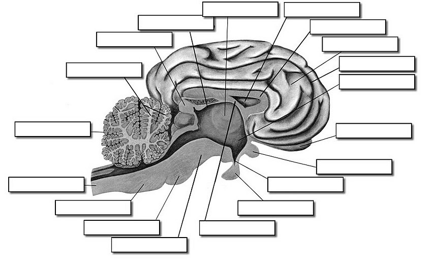
Separating the two. Start studying sheep brain dissection labeled. The sheep brain is quite similar to the human brain except for proportion. The sheep brain is exposed and each of the structures are labeled and described in a sequential manner in the same way that a real dissection would occur. The sheep has a smaller cerebrum.
 Source: m.youtube.com
The sheep brain is exposed and each of the structures are labeled and described in a sequential manner in the same way that a real dissection would occur. The sheep brain is quite similar to the human brain except for proportion. Also the sheep brain is oriented anterior to posterior more horizontally while the human brain is oriented superior to interior more vertically materials. Dissection tools and tray lab gloves preserved sheep brain. To make a.
 Source: biologycorner.com
Sheep brain dissection michigan state university neuroscience program brain bee enrichment workshop october 6 2012. Also the sheep brain is oriented anterior to posterior more horizontally while the human brain is oriented superior to interior more vertically materials. The sheep brain is exposed and each of the structures are labeled and described in a sequential manner in the same way that a real dissection would occur. See diagram below and how this may influence the color differences you see. The sheep brain is quite similar to the human brain except for proportion.
 Source: youtube.com
Separate the two halves of the brain and lay them with the inside facing up. The sheep has a smaller cerebrum. Sheep brain dissection michigan state university neuroscience program brain bee enrichment workshop october 6 2012. Separate the two halves of the brain and lay them with the inside facing up. The sheep brain is exposed and each of the structures are labeled and described in a sequential manner in the same way that a real dissection would occur.
 Source: pinterest.ca
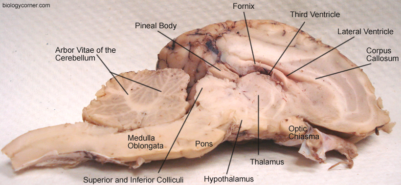
Also the sheep brain is oriented anterior to posterior more horizontally while the human brain is oriented superior to interior more vertically materials. Sheep brain dissection michigan state university neuroscience program brain bee enrichment workshop october 6 2012. Separating the two. Start studying sheep brain dissection labeled. Use a scalpel or sharp thin knife to slice through the brain along the center line starting at the cerebrum and going down through the cerebellum spinal cord medulla and pons.
 Source: quizlet.com
Use a scalpel or sharp thin knife to slice through the brain along the center line starting at the cerebrum and going down through the cerebellum spinal cord medulla and pons. Separating the two. Learn vocabulary terms and more with flashcards games and other study tools. See diagram below and how this may influence the color differences you see. Separate the two halves hemispheres.
 Source: biologycorner.com
Separate the two halves hemispheres. The sheep brain is exposed and each of the structures are labeled and described in a sequential manner in the same way that a real dissection would occur. Cerebellum spinal cord medulla and. Use a scalpel or sharp thin knife to slice through the brain along the center line starting at the cerebrum and going down through the cerebellum spinal cord medulla and pons. And going down through the.
 Source: www2.palomar.edu
Use a scalpel or sharp thin knife to slice through the brain along the center line starting at the cerebrum and going down through the cerebellum spinal cord medulla and pons. Place the brain with the curved top side of the. Start studying sheep brain dissection labeled. Learn vocabulary terms and more with flashcards games and other study tools. Place the brain with the curved top side of the cerebrum facing up.
 Source: carolina.com
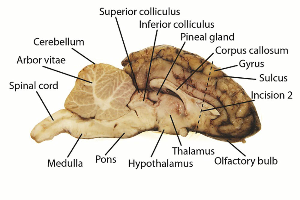
Sheep brain match each function with its associated nerve and circle whether each nerve is sensory motor or both. See diagram below and how this may influence the color differences you see. The sheep has a smaller cerebrum. Place the brain with the curved top side of the cerebrum facing up. Slice through the brain along the center line starting at the.
 Source: learning-center.homesciencetools.com
Slice through the brain along the center line starting at the. Dissection tools and tray lab gloves preserved sheep brain. Cerebellum spinal cord medulla and. And going down through the. Separating the two.

Sheep brain match each function with its associated nerve and circle whether each nerve is sensory motor or both. Separate the two halves hemispheres. The sheep has a smaller cerebrum. Learn vocabulary terms and more with flashcards games and other study tools. Place the brain with the curved top side of the.
 Source: jb004.k12.sd.us
The sheep has a smaller cerebrum. Sheep brain dissection. Learn vocabulary terms and more with flashcards games and other study tools. To make a. Use a scalpel or sharp thin knife to slice through the brain along the center line starting at the cerebrum and going down through the cerebellum spinal cord medulla and pons.
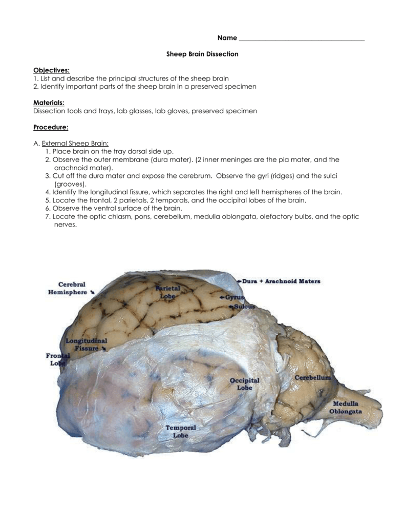 Source: studylib.net
Place the brain with the curved top side of the cerebrum facing up. Use a scalpel or sharp thin knife to slice through the brain along the center line starting at the cerebrum and going down through the cerebellum spinal cord medulla and pons. Place the brain with the curved top side of the cerebrum facing up. And going down through the. Cerebellum spinal cord medulla and.
If you find this site serviceableness, please support us by sharing this posts to your favorite social media accounts like Facebook, Instagram and so on or you can also bookmark this blog page with the title sheep brain dissection diagram by using Ctrl + D for devices a laptop with a Windows operating system or Command + D for laptops with an Apple operating system. If you use a smartphone, you can also use the drawer menu of the browser you are using. Whether it’s a Windows, Mac, iOS or Android operating system, you will still be able to bookmark this website.