Sheep brain dissection labeled
Home » Science Education » Sheep brain dissection labeledSheep brain dissection labeled
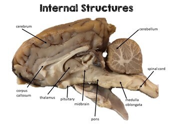
Sheep Brain Dissection Labeled. The sheep has a smaller cerebrum. Muscles other nerves and fatty. Can you tell the difference between the cerebrum and the cerebellum. The anatomy of memory.
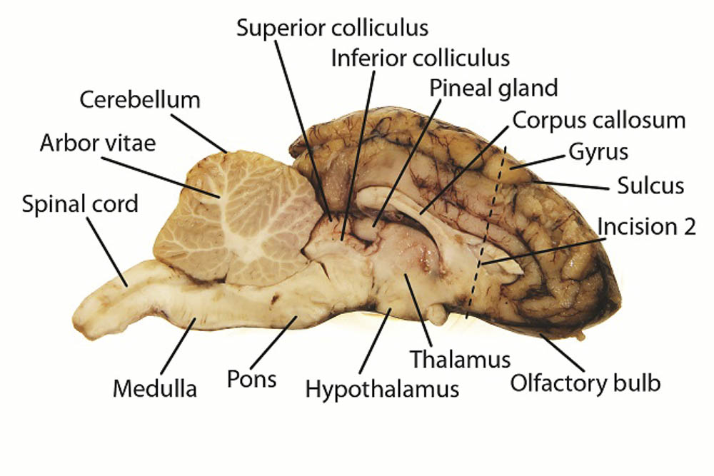 Sheep Brain Dissection Project Guide Hst Learning Center From learning-center.homesciencetools.com
Sheep brain dissection with labeled images sheep brain dissection. A pair of olfactory bulbs may be seen one under each lobe of the frontal cortex. Can you tell the difference between the cerebrum and the cerebellum. The sheep brain is quite similar to the human brain except for proportion. Dissection tools and tray lab gloves preserved sheep brain. The sheep brain is exposed and each of the structures are labeled and described in a sequential manner in the same way that a real dissection would occur.
1 sheep brain and cow eye dissection lab report ivy tech anatomy and physiology 101 2 27 2020 abstract the purpose of the sheep brain and cow eye dissection is to familiarize locating and identify the regions and structures in the brain and eye.
Learn vocabulary terms and more with flashcards games and other study tools. Use a scalpel or sharp thin knife to slice through the brain along the center line starting at the cerebrum and going down through the cerebellum spinal cord medulla and pons. Learn vocabulary terms and more with flashcards games and other study tools. The sheep brain is quite similar to the human brain except for proportion. You ll need a preserved sheep brain for the dissection. Place the brain with the curved top side of the cerebrum facing up.
 Source: starpathdesign.com
The sheep brain is exposed and each of the structures are labeled and described in a sequential manner in the same way that a real dissection would occur. Learn vocabulary terms and more with flashcards games and other study tools. Muscles other nerves and fatty. Start studying sheep brain dissection labeled. 1 sheep brain and cow eye dissection lab report ivy tech anatomy and physiology 101 2 27 2020 abstract the purpose of the sheep brain and cow eye dissection is to familiarize locating and identify the regions and structures in the brain and eye.
 Source: jb004.k12.sd.us
The next several steps will view this surface of the brain. Several important parts of the visual system are visible in the ventral view of the brain. Can you tell the difference between the cerebrum and the cerebellum. You ll need a preserved sheep brain for the dissection. Sheep brain dissection with labeled images sheep brain dissection.
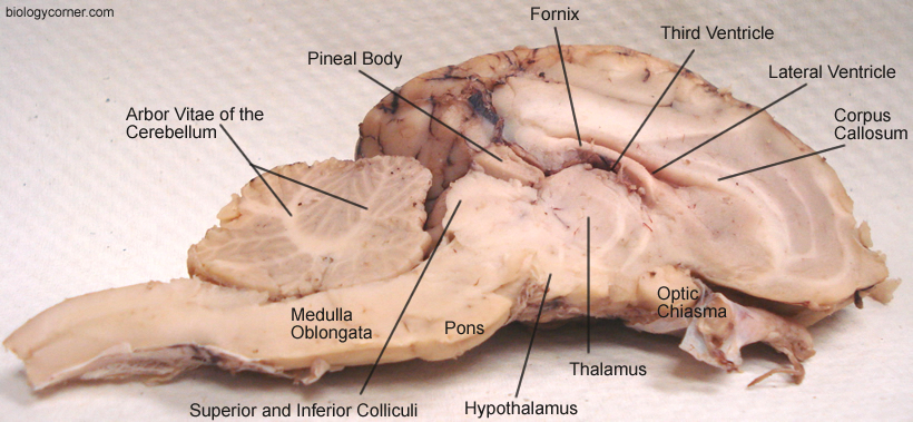 Source: biologycorner.com
By dissecting and labeling the sheep brain and cow. Notice that the brain has two halves or hemispheres. Place the brain with the curved top side of the cerebrum facing up. You ll need a preserved sheep brain for the dissection. Set the brain down so the flatter side with the white spinal cord at one end rests on the dissection pan.
 Source: quizlet.com
The next several steps will view this surface of the brain. Muscles other nerves and fatty. Use a scalpel or sharp thin knife to slice through the brain along the center line starting at the cerebrum and going down through the cerebellum spinal cord medulla and pons. The anatomy of memory. You ll need a preserved sheep brain for the dissection.
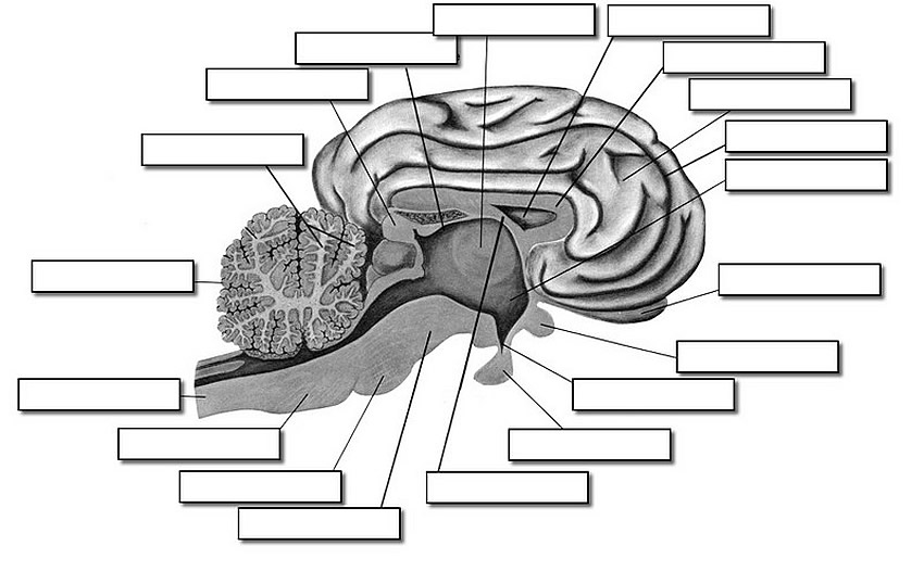 Source: biologycorner.com
By dissecting and labeling the sheep brain and cow. The sheep has a smaller cerebrum. Several important parts of the visual system are visible in the ventral view of the brain. Can you tell the difference between the cerebrum and the cerebellum. Notice that the brain has two halves or hemispheres.
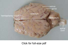 Source: learning-center.homesciencetools.com
The next several steps will view this surface of the brain. Separate the two halves of the brain and lay them with the inside facing up. Set the brain down so the flatter side with the white spinal cord at one end rests on the dissection pan. Sheep brain dissection guide 3. Start studying sheep brain dissection labeled.
 Source: m.youtube.com
Examine the ventral surface of the sheep brain. The anatomy of memory. Separate the two halves of the brain and lay them with the inside facing up. Also the sheep brain is oriented anterior to posterior more horizontally while the human brain is oriented superior to interior more vertically materials. Use a scalpel or sharp thin knife to slice through the brain along the center line starting at the cerebrum and going down through the cerebellum spinal cord medulla and pons.
 Source: teacherspayteachers.com
Set the brain down so the flatter side with the white spinal cord at one end rests on the dissection pan. 1 sheep brain and cow eye dissection lab report ivy tech anatomy and physiology 101 2 27 2020 abstract the purpose of the sheep brain and cow eye dissection is to familiarize locating and identify the regions and structures in the brain and eye. Several important parts of the visual system are visible in the ventral view of the brain. The next several steps will view this surface of the brain. The sheep brain and cow eye were used because their functions are similar of a human brain and eye.

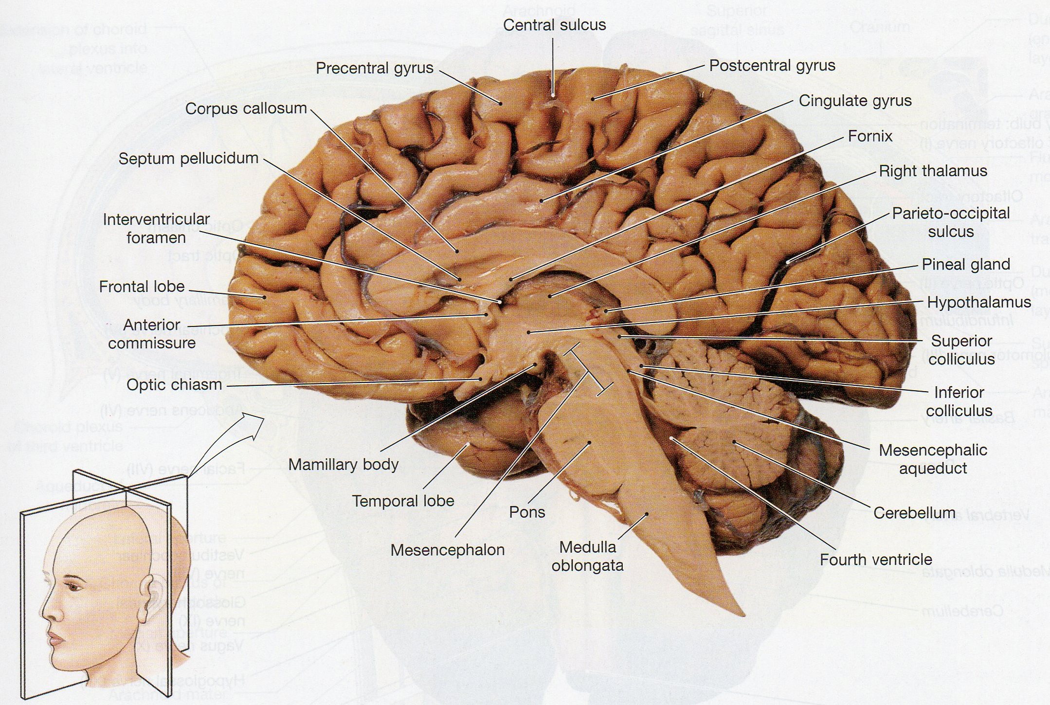
The sheep brain is quite similar to the human brain except for proportion. Several important parts of the visual system are visible in the ventral view of the brain. The sheep brain is exposed and each of the structures are labeled and described in a sequential manner in the same way that a real dissection would occur. Also the sheep brain is oriented anterior to posterior more horizontally while the human brain is oriented superior to interior more vertically materials. Sheep brain dissection guide 3.
 Source: learning-center.homesciencetools.com
Also the sheep brain is oriented anterior to posterior more horizontally while the human brain is oriented superior to interior more vertically materials. Learn vocabulary terms and more with flashcards games and other study tools. The anatomy of memory. Can you tell the difference between the cerebrum and the cerebellum. Notice that the brain has two halves or hemispheres.
 Source: carolina.com
Notice that the brain has two halves or hemispheres. You ll need a preserved sheep brain for the dissection. The exploratorium presents a visual tour of a brain dissection. Use a scalpel or sharp thin knife to slice through the brain along the center line starting at the cerebrum and going down through the cerebellum spinal cord medulla and pons. The next several steps will view this surface of the brain.
 Source: pinterest.ca
Notice that the brain has two halves or hemispheres. Separate the two halves of the brain and lay them with the inside facing up. Set the brain down so the flatter side with the white spinal cord at one end rests on the dissection pan. The next several steps will view this surface of the brain. Start studying sheep brain dissection labeled.
 Source: www2.palomar.edu
Place the brain with the curved top side of the cerebrum facing up. 1 sheep brain and cow eye dissection lab report ivy tech anatomy and physiology 101 2 27 2020 abstract the purpose of the sheep brain and cow eye dissection is to familiarize locating and identify the regions and structures in the brain and eye. Notice that the brain has two halves or hemispheres. The anatomy of memory. Sheep brain dissection guide 3.
 Source: youtube.com
Also the sheep brain is oriented anterior to posterior more horizontally while the human brain is oriented superior to interior more vertically materials. The anatomy of memory. Examine the ventral surface of the sheep brain. Use a scalpel or sharp thin knife to slice through the brain along the center line starting at the cerebrum and going down through the cerebellum spinal cord medulla and pons. Place the brain with the curved top side of the cerebrum facing up.
 Source: courses.lumenlearning.com
Also the sheep brain is oriented anterior to posterior more horizontally while the human brain is oriented superior to interior more vertically materials. The exploratorium presents a visual tour of a brain dissection. The sheep brain and cow eye were used because their functions are similar of a human brain and eye. Examine the ventral surface of the sheep brain. The sheep brain is quite similar to the human brain except for proportion.
If you find this site value, please support us by sharing this posts to your own social media accounts like Facebook, Instagram and so on or you can also bookmark this blog page with the title sheep brain dissection labeled by using Ctrl + D for devices a laptop with a Windows operating system or Command + D for laptops with an Apple operating system. If you use a smartphone, you can also use the drawer menu of the browser you are using. Whether it’s a Windows, Mac, iOS or Android operating system, you will still be able to bookmark this website.
