Sheep eye dissection lab
Home » Science Education » Sheep eye dissection labSheep eye dissection lab
Sheep Eye Dissection Lab. Jomille jasmine kurt joseph 8c by this point we have spotted the aqueous humour. Both my partner and i have learned the structures of the eye. I d heard stories from my mothe r and father both biologists about their cow sheep eye dissections and so i thought i pretty much knew what to expect. The sheep brain and cow eye were used because their functions are similar of a human brain and eye.
 Sheep Eye Dissection From slideshare.net
What is the job of the muscles on the outside of the eyeball. Sheep eye virtual dissection lab 35 part a discussion 29 marks 1. The sheep eye has a rectangular pupil shape. The sheep brain and cow eye were used because their functions are similar of a human brain and eye. I d heard stories from my mothe r and father both biologists about their cow sheep eye dissections and so i thought i pretty much knew what to expect. The four extrinsic muscles humans have six move the sheep eye while the fatty tissue cushions the eye.
What colour is the.
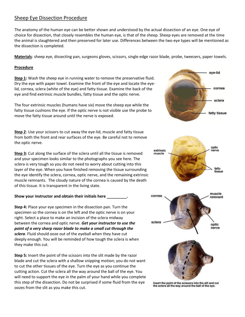
Wash the sheep eye in running water to remove the preservative fluid. 2 the job of the muscles on the outside of the eyeball also known as the extrinsic or extraocular muscles is to control and move the eyeball in different directions through the contraction of the muscles. I d heard stories from my mothe r and father both biologists about their cow sheep eye dissections and so i thought i pretty much knew what to expect. The sheep brain and cow eye were used because their functions are similar of a human brain and eye. Sheep eye dissection analysis in this lab we dissected a sheep s eye. 1 sheep brain and cow eye dissection lab report ivy tech anatomy and physiology 101 2 27 2020 abstract the purpose of the sheep brain and cow eye dissection is to familiarize locating and identify the regions and structures in the brain and eye.
 Source: m.youtube.com
What colour is the. What is the job of the muscles on the outside of the eyeball. This is after making an incision on the thin layer of the cornea. Sheep eye dissection analysis in this lab we dissected a sheep s eye. For example the iris.
 Source: sites.google.com
Not only did we learn the structure of the eye but also the function that each part serves. Wash the sheep eye in running water to remove the preservative fluid. For example the iris. Most of the things that i d heard about did happen the black liquid squishing forth once the cornea had been punctured the little gooey jelly bag inside. Dry the eye with paper toweling.
 Source: studylib.net
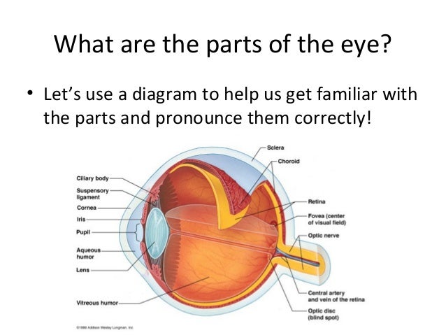
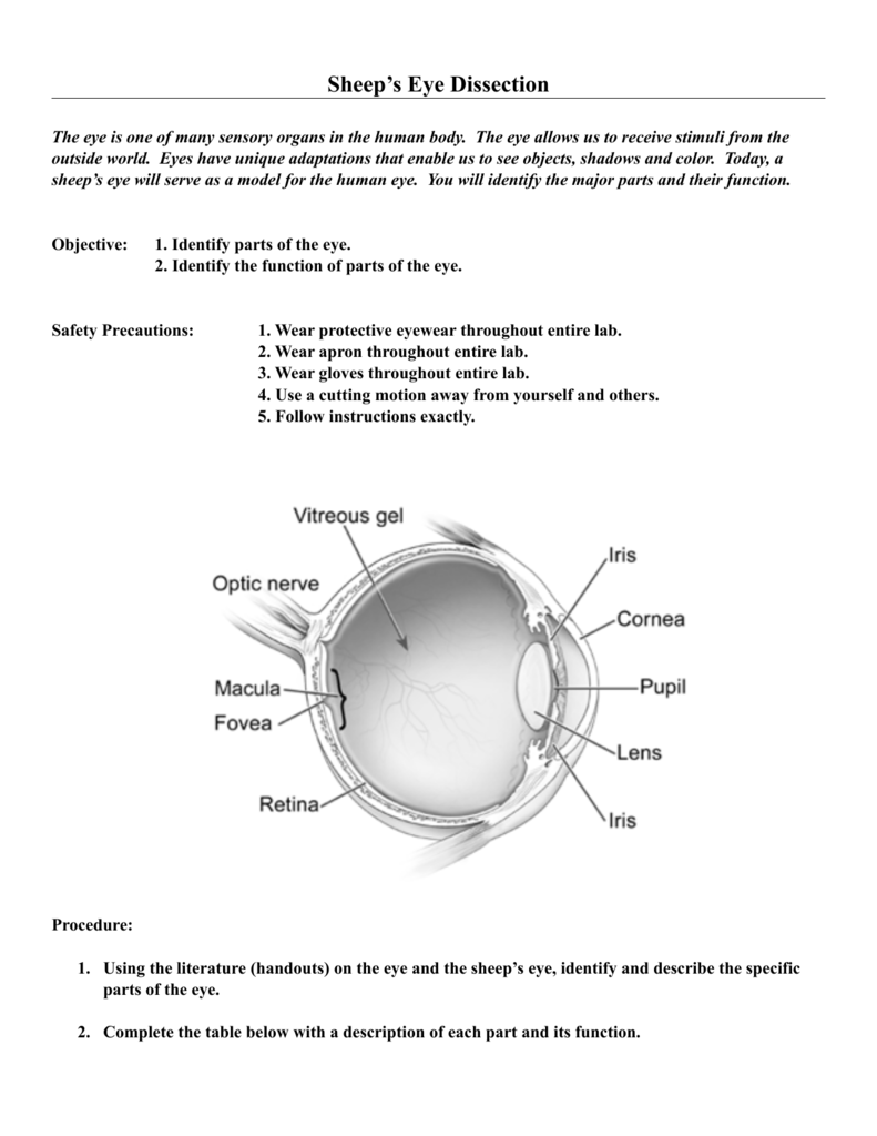
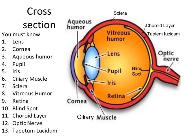
A sheep s eye is very similar to our own except they need to see in low light environments meaning that they have a tapetum lucidum on the choroid coat that allows light to reflect more onto the retina. Examine the back of the eye and find extrinsic muscle bundles fatty tissue and the optic nerve. A sheep s eye is very similar to our own except they need to see in low light environments meaning that they have a tapetum lucidum on the choroid coat that allows light to reflect more onto the retina. The function of the aqueous humour is to not only moisturize the. What colour is the.
 Source: slideshare.net
Jomille jasmine kurt joseph 8c by this point we have spotted the aqueous humour. For example the iris. Dry the eye with paper toweling. What is the job of the muscles on the outside of the eyeball. 1 sheep brain and cow eye dissection lab report ivy tech anatomy and physiology 101 2 27 2020 abstract the purpose of the sheep brain and cow eye dissection is to familiarize locating and identify the regions and structures in the brain and eye.
 Source: youtube.com
Wash the sheep eye in running water to remove the preservative fluid. The sheep brain and cow eye were used because their functions are similar of a human brain and eye. Not only did we learn the structure of the eye but also the function that each part serves. The four extrinsic muscles humans have six move the sheep eye while the fatty tissue cushions the eye. This is after making an incision on the thin layer of the cornea.
 Source: quizlet.com
A sheep s eye is very similar to our own except they need to see in low light environments meaning that they have a tapetum lucidum on the choroid coat that allows light to reflect more onto the retina. Most of the things that i d heard about did happen the black liquid squishing forth once the cornea had been punctured the little gooey jelly bag inside. Sheep eye virtual dissection lab 35 part a discussion 29 marks 1. Sheep eye dissection analysis in this lab we dissected a sheep s eye. These structures include the sclera vitreous humor retina choroid coat lens ciliary body etc.
 Source: pinterest.com
On thursday the 17th my biology class did an interesting lab. Examine the back of the eye and find extrinsic muscle bundles fatty tissue and the optic nerve. A sheep s eye is very similar to our own except they need to see in low light environments meaning that they have a tapetum lucidum on the choroid coat that allows light to reflect more onto the retina. These structures include the sclera vitreous humor retina choroid coat lens ciliary body etc. Most of the things that i d heard about did happen the black liquid squishing forth once the cornea had been punctured the little gooey jelly bag inside.

Examine the back of the eye and find extrinsic muscle bundles fatty tissue and the optic nerve. Dry the eye with paper toweling. By dissecting and labeling the sheep brain and cow eye gives hands on experience and help memorize location of each structure. This is after making an incision on the thin layer of the cornea. I d heard stories from my mothe r and father both biologists about their cow sheep eye dissections and so i thought i pretty much knew what to expect.
 Source: studylib.net
Dry the eye with paper toweling. The function of the aqueous humour is to not only moisturize the. If the optic nerve is not visible use the probe to move the fatty tissue. On thursday the 17th my biology class did an interesting lab. Both my partner and i have learned the structures of the eye.
 Source: amazon.com
For example the iris. 2 the job of the muscles on the outside of the eyeball also known as the extrinsic or extraocular muscles is to control and move the eyeball in different directions through the contraction of the muscles. Most of the things that i d heard about did happen the black liquid squishing forth once the cornea had been punctured the little gooey jelly bag inside. Dry the eye with paper toweling. A sheep s eye is very similar to our own except they need to see in low light environments meaning that they have a tapetum lucidum on the choroid coat that allows light to reflect more onto the retina.
 Source: slideshare.net
Sheep eye dissection analysis in this lab we dissected a sheep s eye. These structures include the sclera vitreous humor retina choroid coat lens ciliary body etc. I d heard stories from my mothe r and father both biologists about their cow sheep eye dissections and so i thought i pretty much knew what to expect. This is after making an incision on the thin layer of the cornea. If the optic nerve is not visible use the probe to move the fatty tissue.
 Source: science.jburroughs.org
These structures include the sclera vitreous humor retina choroid coat lens ciliary body etc. These structures include the sclera vitreous humor retina choroid coat lens ciliary body etc. The function of the aqueous humour is to not only moisturize the. Sheep eye dissection analysis in this lab we dissected a sheep s eye. 2 the job of the muscles on the outside of the eyeball also known as the extrinsic or extraocular muscles is to control and move the eyeball in different directions through the contraction of the muscles.
 Source: studylib.net
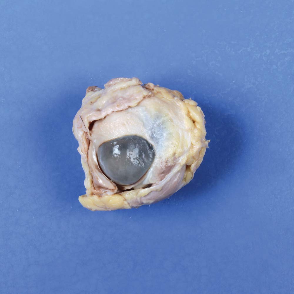
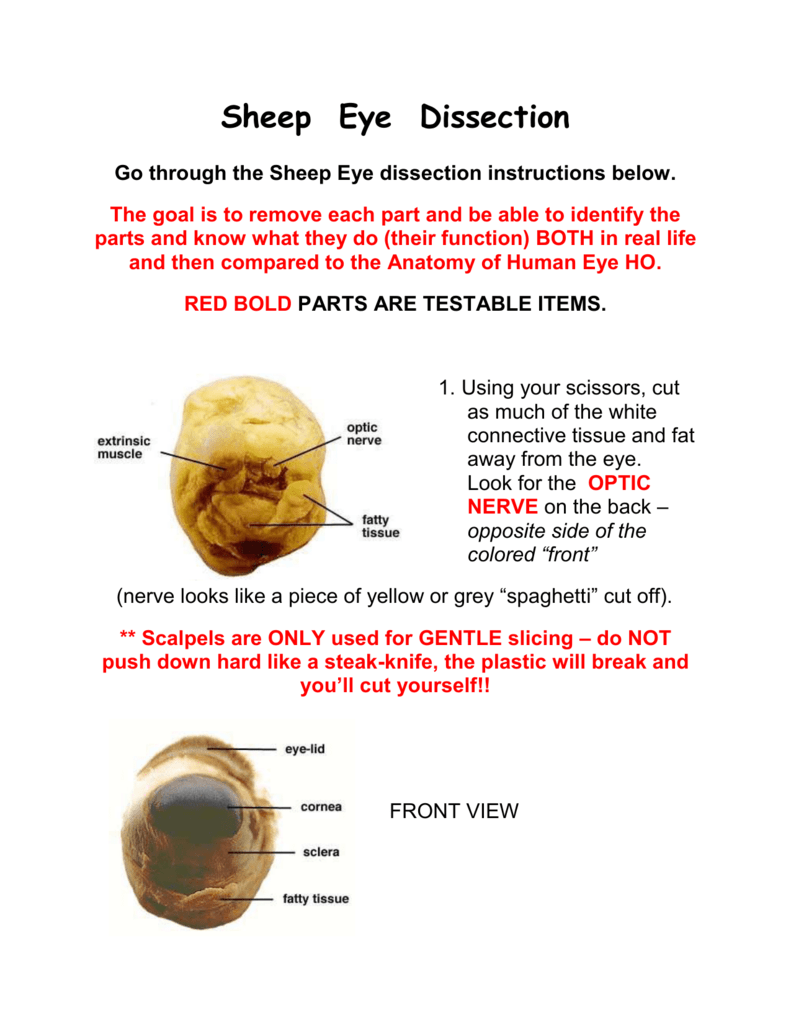
Both my partner and i have learned the structures of the eye. Both my partner and i have learned the structures of the eye. What colour is the. Examine the front of the eye and locate the eye lid cornea sclera white of the eye and fatty tissue. I d heard stories from my mothe r and father both biologists about their cow sheep eye dissections and so i thought i pretty much knew what to expect.
 Source: mohtadialkhaliq.wordpress.com
If the optic nerve is not visible use the probe to move the fatty tissue. The function of the aqueous humour is to not only moisturize the. On thursday the 17th my biology class did an interesting lab. Examine the back of the eye and find extrinsic muscle bundles fatty tissue and the optic nerve. The four extrinsic muscles humans have six move the sheep eye while the fatty tissue cushions the eye.
 Source: science.jburroughs.org
Jomille jasmine kurt joseph 8c by this point we have spotted the aqueous humour. The four extrinsic muscles humans have six move the sheep eye while the fatty tissue cushions the eye. Wash the sheep eye in running water to remove the preservative fluid. If the optic nerve is not visible use the probe to move the fatty tissue. Examine the back of the eye and find extrinsic muscle bundles fatty tissue and the optic nerve.
If you find this site serviceableness, please support us by sharing this posts to your preference social media accounts like Facebook, Instagram and so on or you can also bookmark this blog page with the title sheep eye dissection lab by using Ctrl + D for devices a laptop with a Windows operating system or Command + D for laptops with an Apple operating system. If you use a smartphone, you can also use the drawer menu of the browser you are using. Whether it’s a Windows, Mac, iOS or Android operating system, you will still be able to bookmark this website.
