Sheep heart anatomy labeled
Home » Science Education » Sheep heart anatomy labeledSheep heart anatomy labeled
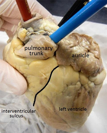
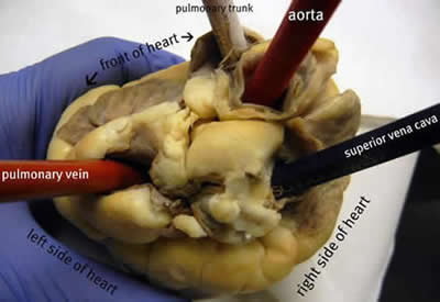
Sheep Heart Anatomy Labeled. Label the parts of a sheep heart. All the major vessels are represented many are labeled with colored pencils so that you can see. Use a scalpel to make an incision in the heart at the superior vena cava. Labeled sheep heart picture 1.

Made with explain everything. Other sets by this creator. Left and right ventricle left and right atrium pulmonary artery pulmonary veins aorta coronary artery apex superior and inferior vena cava. Use a scalpel to make an incision in the heart at the superior vena cava. Blood from the tissues superior and inferior vena cava right atrium tricuspid valve right ventricle pulmonary semilunar valve pulmonary artery lungs pulmonary veins left atrium bicuspid mitral valve left ventricle aortic semilunar valve aorta body tissue. Jun 12 2016 this page contains photos of the sheep heart dissection.
Human anatomy and physiology 2 sheep heart anatomy demonstration.
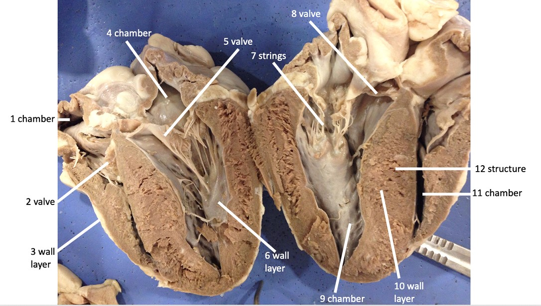
Labeled sheep heart picture 1. Locate the pulmonary. The incision should follow the line of the right side of the heart so that you can open just the right side and see the right atrium the right ventricle and the tricuspid valve between them. Left and right ventricle left and right atrium pulmonary artery pulmonary veins aorta coronary artery apex superior and inferior vena cava. Study materials teaching materials cardiac cycle nurse aesthetic heart anatomy medical anatomy human anatomy and physiology anatomy study heart pictures. Blood from the tissues superior and inferior vena cava right atrium tricuspid valve right ventricle pulmonary semilunar valve pulmonary artery lungs pulmonary veins left atrium bicuspid mitral valve left ventricle aortic semilunar valve aorta body tissue.
Source: sen842cova.blogspot.com
Article by minted peach. Label the parts of a sheep heart. Article by minted peach. The heart can be confusing because it is not perfectly symmetrical. Human anatomy and physiology 2 sheep heart anatomy demonstration.
 Source: br.pinterest.com
All the major vessels are represented many are labeled with colored pencils so that you can see. The heart can be confusing because it is not perfectly symmetrical. This page contains photos of the sheep heart dissection. Heart anatomy lab exam 1. Label the parts of a sheep heart.
 Source: pinterest.cl
The incision should follow the line of the right side of the heart so that you can open just the right side and see the right atrium the right ventricle and the tricuspid valve between them. Made with explain everything. Heart anatomy lab exam 1. Study materials teaching materials cardiac cycle nurse aesthetic heart anatomy medical anatomy human anatomy and physiology anatomy study heart pictures. Labeled sheep heart picture 1.
 Source: youtube.com
Locate the pulmonary. Heart anatomy lab exam 1. The heart can be confusing because it is not perfectly symmetrical. Nicellian has uploaded 157 photos to flickr. All the major vessels are represented many are labeled with colored pencils so that you can see exactly where each is located.

This page contains photos of the sheep heart dissection. All the major vessels are represented many are labeled with colored pencils so that you can see. Reed section 1 open water diver manual notes. Label the parts of a sheep heart. Sheep heart anatomy including atria ventricles vena cava aorta pulmonary artery vein tricuspid valve bicuspid valve aortic and pulmonic valves trab.
 Source: biologycorner.com
Left and right ventricle left and right atrium pulmonary artery pulmonary veins aorta coronary artery apex superior and inferior vena cava. Article by minted peach. All the major vessels are represented many are labeled with colored pencils so that you can see exactly where each is located. Jun 12 2016 this page contains photos of the sheep heart dissection. All the major vessels are represented many are labeled with colored pencils so that you can see.

Study materials teaching materials cardiac cycle nurse aesthetic heart anatomy medical anatomy human anatomy and physiology anatomy study heart pictures. Explore nicellian s photos on flickr. Made with explain everything. The incision should follow the line of the right side of the heart so that you can open just the right side and see the right atrium the right ventricle and the tricuspid valve between them. Sheep heart dissection procedure day 2 you will be cutting the heart open today.
 Source: anatomycorner.com
Study materials teaching materials cardiac cycle nurse aesthetic heart anatomy medical anatomy human anatomy and physiology anatomy study heart pictures. The incision should follow the line of the right side of the heart so that you can open just the right side and see the right atrium the right ventricle and the tricuspid valve between them. This page contains photos of the sheep heart dissection. Sheep heart anatomy including atria ventricles vena cava aorta pulmonary artery vein tricuspid valve bicuspid valve aortic and pulmonic valves trab. All the major vessels are represented many are labeled with colored pencils so that you can see.
 Source: quizlet.com
Heart anatomy lab exam 1. Nicellian has uploaded 157 photos to flickr. Article by minted peach. Other sets by this creator. Students often confuse the left and the right side of the heart.

Use a scalpel to make an incision in the heart at the superior vena cava. Locate the pulmonary. Labeled sheep heart picture 1. Heart anatomy lab exam 1. This page contains photos of the sheep heart dissection.
 Source: quizlet.com
Use a scalpel to make an incision in the heart at the superior vena cava. Label the parts of a sheep heart. Article by minted peach. Study materials teaching materials cardiac cycle nurse aesthetic heart anatomy medical anatomy human anatomy and physiology anatomy study heart pictures. Blood from the tissues superior and inferior vena cava right atrium tricuspid valve right ventricle pulmonary semilunar valve pulmonary artery lungs pulmonary veins left atrium bicuspid mitral valve left ventricle aortic semilunar valve aorta body tissue.
 Source: chegg.com
Made with explain everything. Explore nicellian s photos on flickr. Jun 12 2016 this page contains photos of the sheep heart dissection. All the major vessels are represented many are labeled with colored pencils so that you can see exactly where each is located. Sheep heart dissection procedure day 2 you will be cutting the heart open today.
 Source: pinterest.com
Reed section 1 open water diver manual notes. The incision should follow the line of the right side of the heart so that you can open just the right side and see the right atrium the right ventricle and the tricuspid valve between them. Article by minted peach. Heart anatomy lab exam 1. Other sets by this creator.
 Source: biologycorner.com
All the major vessels are represented many are labeled with colored pencils so that you can see. Locate the pulmonary. Nicellian has uploaded 157 photos to flickr. Students often confuse the left and the right side of the heart. Article by minted peach.
 Source: pinterest.com
Explore nicellian s photos on flickr. Review the outer part of the heart and make sure you know where these structures are. Locate the pulmonary. Use a scalpel to make an incision in the heart at the superior vena cava. Sheep heart anatomy including atria ventricles vena cava aorta pulmonary artery vein tricuspid valve bicuspid valve aortic and pulmonic valves trab.
If you find this site beneficial, please support us by sharing this posts to your favorite social media accounts like Facebook, Instagram and so on or you can also save this blog page with the title sheep heart anatomy labeled by using Ctrl + D for devices a laptop with a Windows operating system or Command + D for laptops with an Apple operating system. If you use a smartphone, you can also use the drawer menu of the browser you are using. Whether it’s a Windows, Mac, iOS or Android operating system, you will still be able to bookmark this website.
