Staining gram negative bacteria
Home » Science Education » Staining gram negative bacteriaStaining gram negative bacteria
Staining Gram Negative Bacteria. Gram positive cells have a thick layer of peptidoglycan in the cell wall that retains the primary st. Gram staining differentiates bacteria by the chemical and physical properties of their cell walls. They are characterized by their cell envelopes which are composed of a thin peptidoglycan cell wall sandwiched between an inner cytoplasmic cell membrane and a bacterial outer membrane. Differential stains gram stain.
 Gram Stain Procedure In Microbiology From thoughtco.com
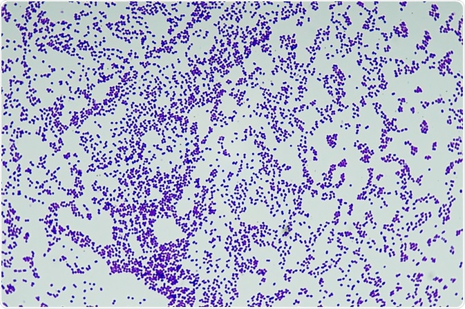
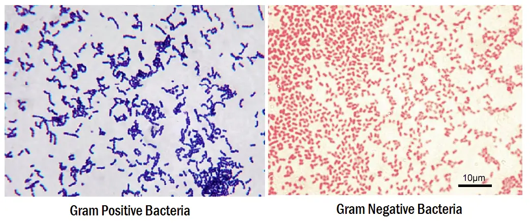
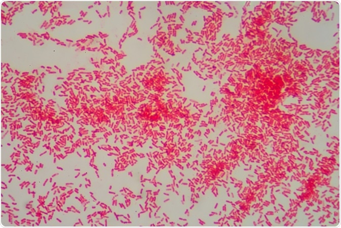
Gram staining a differential staining method. Gram staining differentiates bacteria by the chemical and physical properties of their cell walls. Gram staining procedure is developed in 1884 by hans christian gram. Find information and process for the preparation of gram staining regent. The gram negative bacteria appear colorless and gram positive bacteria remain blue. The red dye safranin stains the decolorized gram negative cells red pink.
Gram staining is the primary step for the initial identification and classification of the bacteria into two groups i e.
Application of counterstain safranin. Gram positive bacteria stain violet due to the presence of a thick layer of peptidoglycan in their cell walls which retains the crystal violet these cells are stained with. The gram negative bacteria appear colorless and gram positive bacteria remain blue. It is used to identify and differentiate bacteria into two groups i e. Gram stain or gram staining also called gram s method is a method of staining used to distinguish and classify bacterial species into two large groups. It stains the bacterial cell wall differently by giving violet colour to the gram positive bacteria and pink colour to the negative bacteria.
 Source: news-medical.net
In a final step a counterstain such as safranin is added and stains the gram negative cells red. Gram staining a differential staining method. Gram staining differentiates bacteria by the chemical and physical properties of their cell walls. In a final step a counterstain such as safranin is added and stains the gram negative cells red. Gram positive cells have a thick layer of peptidoglycan in the cell wall that retains the primary st.
 Source: tmedweb.tulane.edu
One of the most well known differential stains is gram stain which differentiates gram positive and gram negative bacteria based on the difference in their cell wall structure. Application of counterstain safranin. Differential stains gram stain. The gram positive bacteria remain blue. It is also known as gram staining or gram s method.
 Source: thoughtco.com
Gram stain or gram staining also called gram s method is a method of staining used to distinguish and classify bacterial species into two large groups. Application of counterstain safranin. The gram stain was developed by the danish bacteriologist hans christian gram. Differential stains gram stain. Gram staining procedure is developed in 1884 by hans christian gram.
 Source: aatbio.com
Differential stains gram stain. Gram staining procedure is developed in 1884 by hans christian gram. The procedure is named for the person who developed the technique danish bacteriologist hans christian gram. Gram negative bacteria however are decolorized because they have cell walls with much thinner layers that allow removal of the dye by the solvent. The gram stain is a differential method of staining used to assign bacteria to one of two groups gram positive and gram negative based on the properties of their cell walls.
 Source: verywellhealth.com
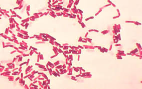
It stains the bacterial cell wall differently by giving violet colour to the gram positive bacteria and pink colour to the negative bacteria. Application of counterstain safranin. The gram stain was developed by the danish bacteriologist hans christian gram. The red dye safranin stains the decolorized gram negative cells red pink. Find information and process for the preparation of gram staining regent.
 Source: news-medical.net
The name comes from the danish bacteriologist hans christian gram who developed the technique. The gram negative bacteria appear colorless and gram positive bacteria remain blue. Differential stains gram stain. The gram positive bacteria remain blue. Gram negative bacteria however are decolorized because they have cell walls with much thinner layers that allow removal of the dye by the solvent.
 Source: en.wikipedia.org
Alternatively gram negative bacteria stain red which is attributed to a thinner peptidoglycan wall which does not retain the crystal violet during the decoloring process. Gram staining a differential staining method. Gram stain or gram staining also called gram s method is a method of staining used to distinguish and classify bacterial species into two large groups. Principle of gram stain image 2. The name comes from the danish bacteriologist hans christian gram who developed the technique.
 Source: microbenotes.com
Gram positive bacteria stain violet due to the presence of a thick layer of peptidoglycan in their cell walls which retains the crystal violet these cells are stained with. One of the most well known differential stains is gram stain which differentiates gram positive and gram negative bacteria based on the difference in their cell wall structure. Gram staining differentiates bacteria by the chemical and physical properties of their cell walls. The procedure is named for the person who developed the technique danish bacteriologist hans christian gram. Gram negative bacteria are bacteria that do not retain the crystal violet stain used in the gram staining method of bacterial differentiation.
 Source: thisonevsthatone.com
In a final step a counterstain such as safranin is added and stains the gram negative cells red. Gram staining differentiates bacteria by the chemical and physical properties of their cell walls. Gram positive bacteria and gram negative bacteria. Gram stain or gram staining also called gram s method is a method of staining used to distinguish and classify bacterial species into two large groups. It is also known as gram staining or gram s method.
 Source: technologynetworks.com
Gram positive bacteria remain purple because they have a single thick cell wall that is not easily penetrated by the solvent. Gram negative bacteria are found everywhere in virtually all. The gram positive bacteria remain blue. Gram staining a differential staining method. The name comes from the danish bacteriologist hans christian gram who developed the technique.
 Source: atsu.edu
Gram positive bacteria and gram negative bacteria. Find information and process for the preparation of gram staining regent. In contrast to simple stains differential stains are used to distinguish the difference between bacteria. The procedure is named for the person who developed the technique danish bacteriologist hans christian gram. Gram negative bacteria are found everywhere in virtually all.
 Source: en.wikipedia.org
Differential stains gram stain. Gram negative bacteria are bacteria that do not retain the crystal violet stain used in the gram staining method of bacterial differentiation. Alternatively gram negative bacteria stain red which is attributed to a thinner peptidoglycan wall which does not retain the crystal violet during the decoloring process. The procedure is named for the person who developed the technique danish bacteriologist hans christian gram. Gram stain or gram staining also called gram s method is a method of staining used to distinguish and classify bacterial species into two large groups.
 Source: ib.bioninja.com.au
Gram staining procedure is developed in 1884 by hans christian gram. Gram negative bacteria are bacteria that do not retain the crystal violet stain used in the gram staining method of bacterial differentiation. Principle of gram stain image 2. Differential stains gram stain. They are characterized by their cell envelopes which are composed of a thin peptidoglycan cell wall sandwiched between an inner cytoplasmic cell membrane and a bacterial outer membrane.
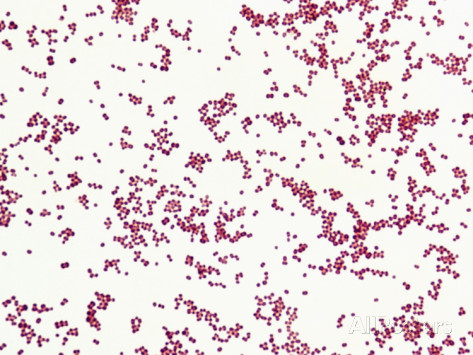 Source: atsu.edu
Gram negative bacteria however are decolorized because they have cell walls with much thinner layers that allow removal of the dye by the solvent. Gram negative bacteria are found everywhere in virtually all. Gram stain or gram staining also called gram s method is a method of staining used to distinguish and classify bacterial species into two large groups. Find information and process for the preparation of gram staining regent. The gram stain is a differential method of staining used to assign bacteria to one of two groups gram positive and gram negative based on the properties of their cell walls.
 Source: microdok.com
In a final step a counterstain such as safranin is added and stains the gram negative cells red. It is also known as gram staining or gram s method. It is used to identify and differentiate bacteria into two groups i e. This classification is based on the physical properties of the bacterial cell wall. Gram negative bacteria are found everywhere in virtually all.
If you find this site helpful, please support us by sharing this posts to your favorite social media accounts like Facebook, Instagram and so on or you can also bookmark this blog page with the title staining gram negative bacteria by using Ctrl + D for devices a laptop with a Windows operating system or Command + D for laptops with an Apple operating system. If you use a smartphone, you can also use the drawer menu of the browser you are using. Whether it’s a Windows, Mac, iOS or Android operating system, you will still be able to bookmark this website.
